Image & data credits
The patient anatomy in the RT Image Matching Trainer — and in the screenshots and animations of it shown on this site — is derived from medical-imaging datasets that researchers and institutions have published under open Creative Commons Attribution (CC BY) licences. This page records the cited source, licence, processing, and known research-participant conditions; an open release or licence does not by itself establish a dataset's privacy, consent, or re-identification status. The two Classroom screenshots are deterministic fictional UI fixtures: their names, class codes, assignments, adjustments, and results are invented, and they contain no patient image.
Educational use only. Every case has been adapted for training and changes were made to all source data: volumes are cropped, resampled, reconstructed, and re-windowed; radiographs and DRRs may be computer-rendered; and several cases add synthetic training targets (lesions, target volumes, plans, or surgical clips) that do not exist in the source scans. Unless a case explicitly identifies released source plan, acquisition, contour, or motion metadata, setup offsets, tolerances, and readouts are fictional. Source treatment metadata are replayed only as a non-clinical teaching simulation; the trainer does not reproduce or validate clinical dose.
Radiation therapy profile artwork
The twelve profile avatars are original procedural Blender miniatures created for this project with Higgsfield's 3D Jutsu tools: Isocentre, Beam aperture, Dose curves, Linac, CT scanner, MLC, Treatment couch, IGRT panel, QA phantom, Thermoplastic mask, Photon beam and Electron cone. They use independently constructed geometry and materials, with no patient images, imported models, textures or equipment photographs. The mask uses an original synthetic head form. These stylized images are decorative profile choices and do not represent clinical geometry, dose or professional qualifications.
Retained landing media
These earlier audited landing-page assets remain available for existing links and capture provenance. The current landing page uses the cinematic captures below.
- machine-tour-mobile-poster-v2.png
- machine-tour-mobile-v2.mp4
- machine-tour-poster-v2.png
- machine-tour-v2.mp4
- scroll-tour-mobile-v2.mp4
- scroll-tour-v2.mp4
- feature-film-v7.mp4
- feature-film-v7-poster.webp
Known research-participant conditions and any source-specific privacy evidence we found are recorded on this page; evidence not documented remains unestablished.
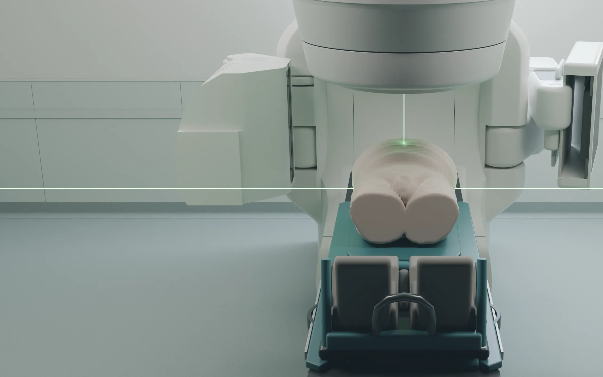
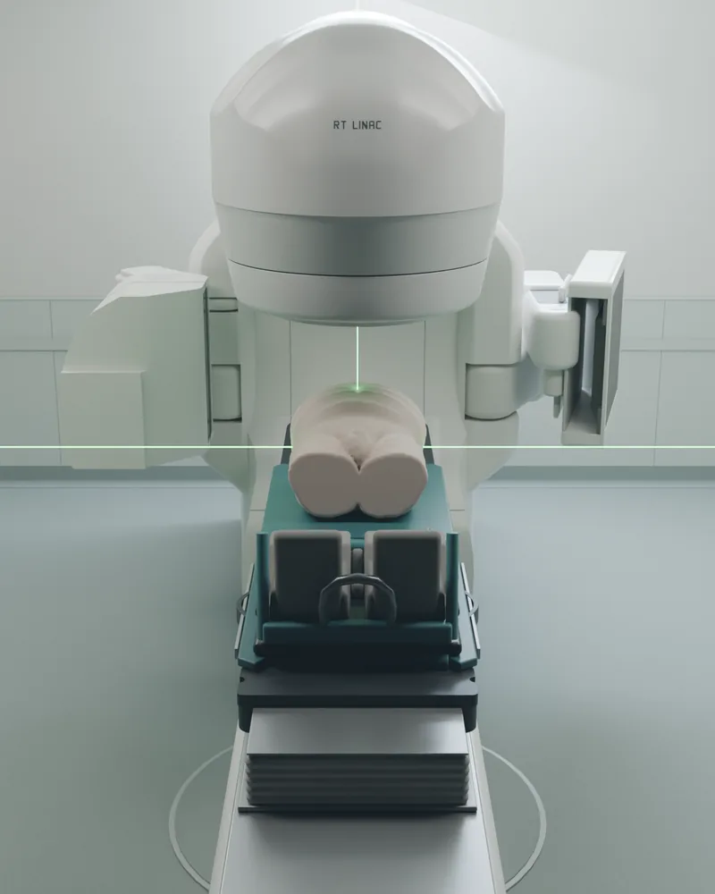
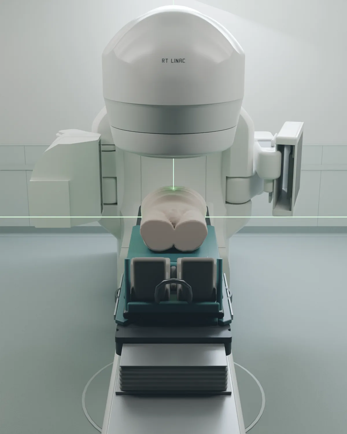
Opening treatment room: October 19 render
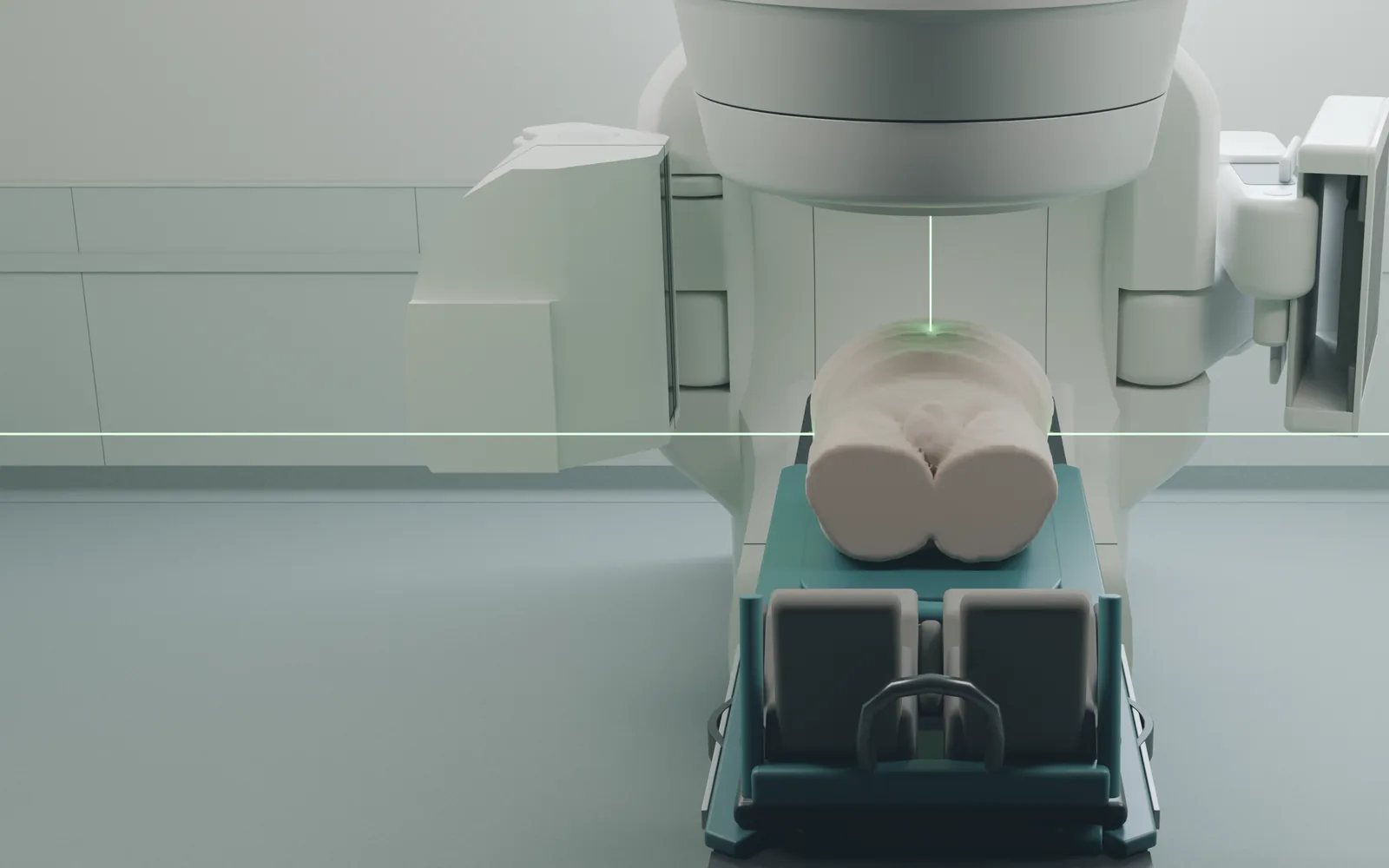
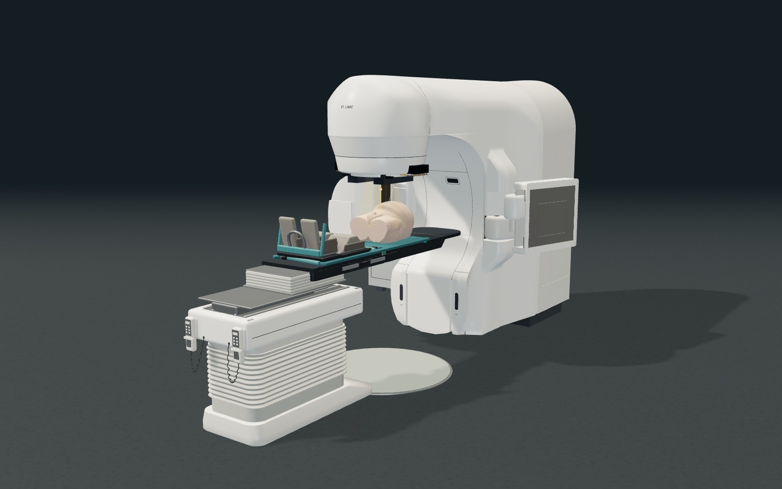
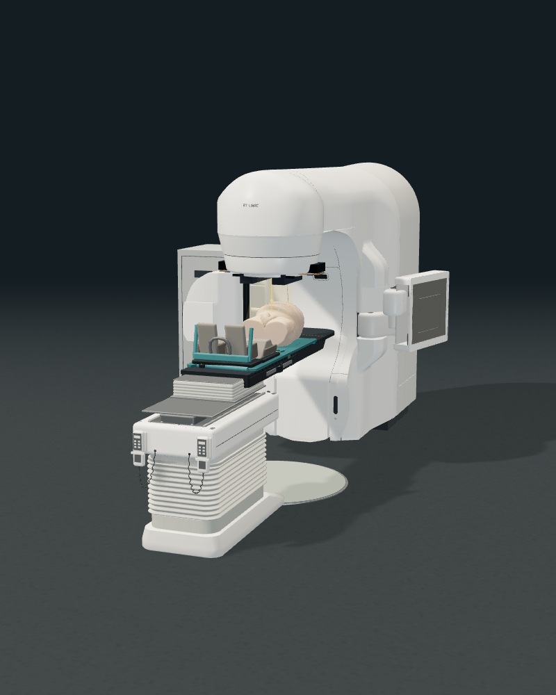
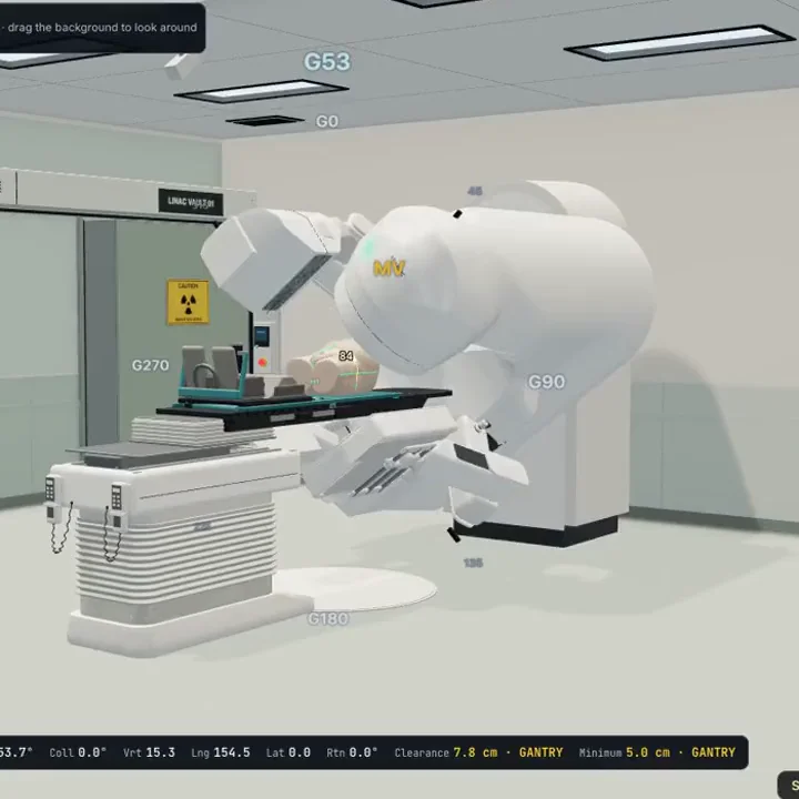
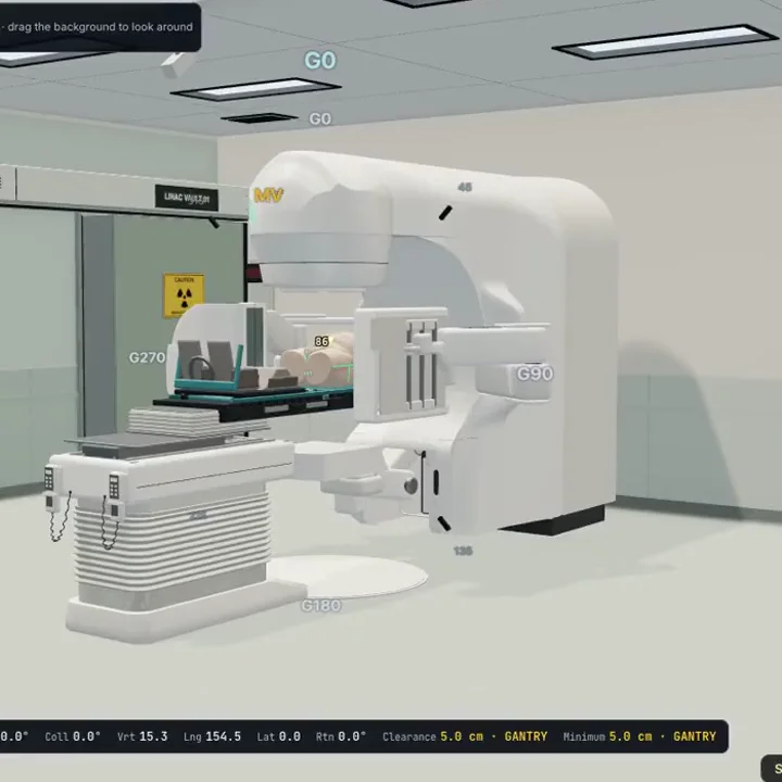
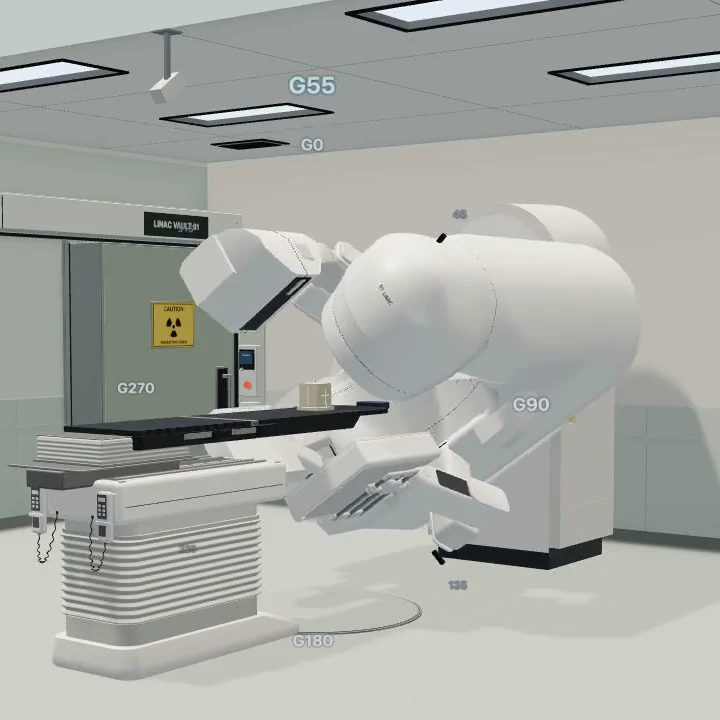
The lit treatment room that opens the landing page is the first frame of the room film, rendered in Blender from the trainer’s own 3D models: its treatment room, linac, couch and Prostate VMAT patient, whose pelvic exterior is derived from Prostate Anatomical Edge Cases (TCIA), CC BY 4.0, with the full citation below, and no head or facial source anatomy. The trainer supplies the geometry and positions; lighting, camera and the rebuilt room lasers are staged. No pixels are generated or retouched. Each layout is rendered once, at 160% (desktop) or 150% (phone) of the size it is laid out at: high-density screens receive that render, and other screens the same render downsampled, so every size shows the same frame. The training equipment is representative geometry, independent of equipment manufacturers. Render inputs, isocentre positions and file hashes are recorded in docs/landing-room-hero.json and the rights register.
- Treatment room, desktop
- Treatment room, desktop, high density
- Treatment room, phone
- Treatment room, phone, high density
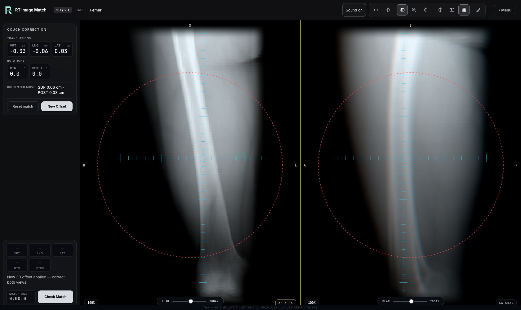
Cinematic design preview: October 9 captures
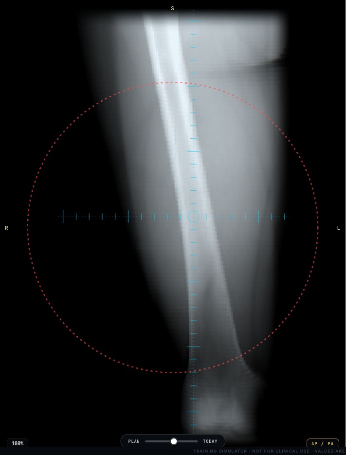
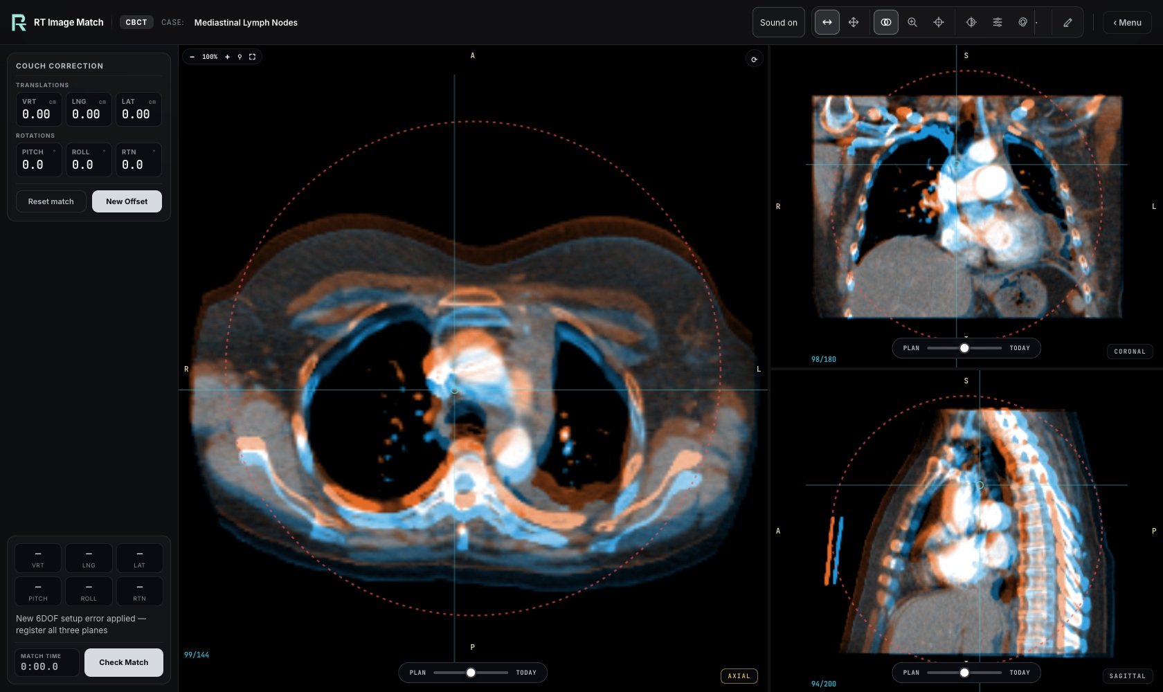
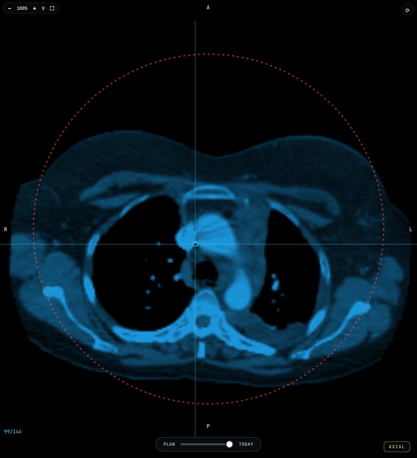
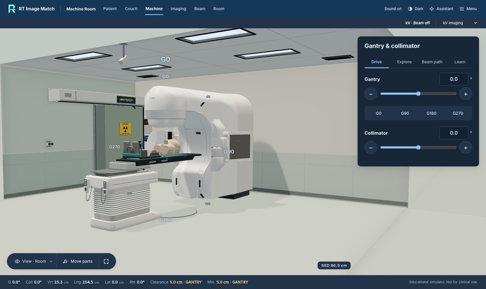
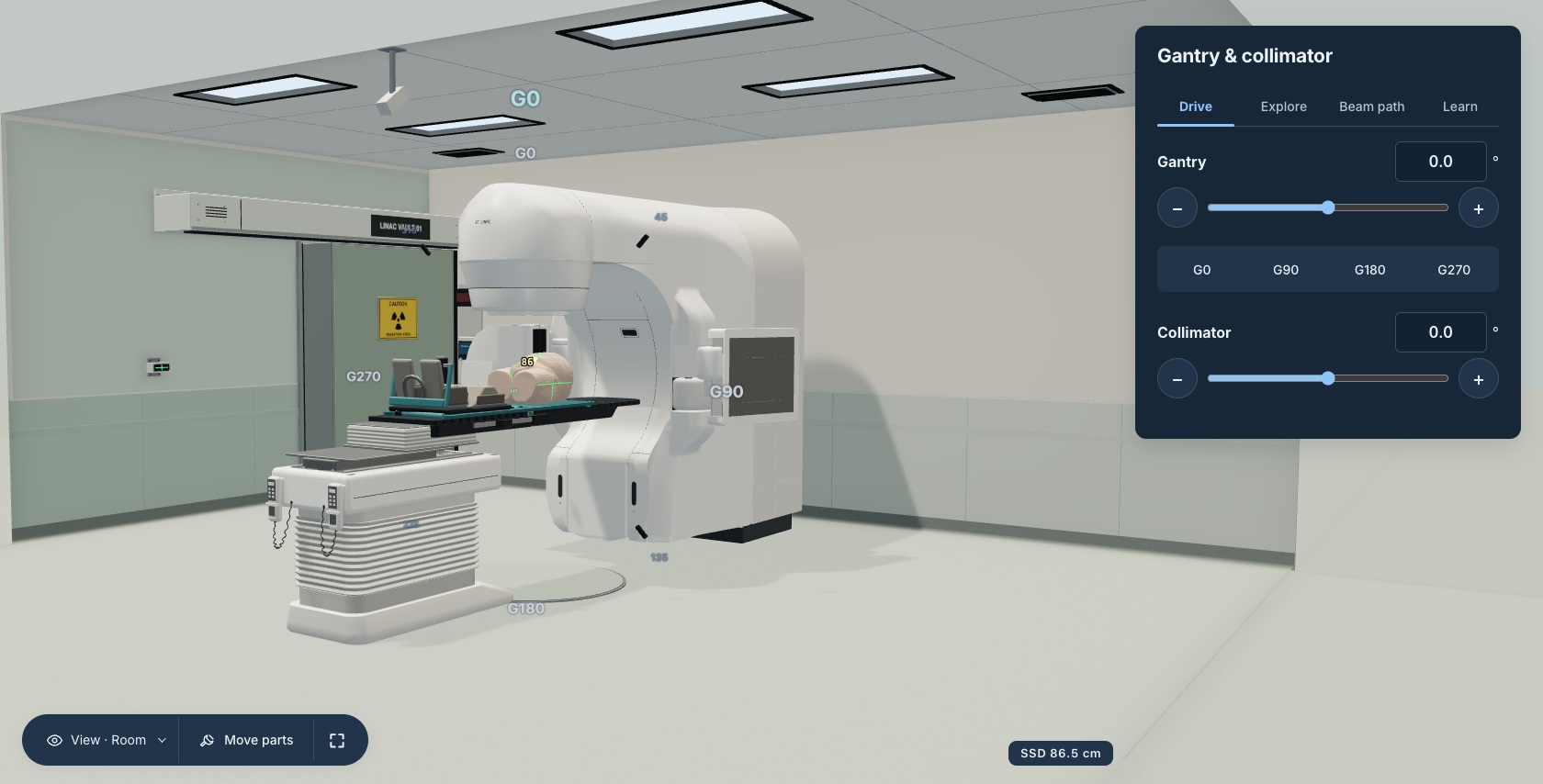
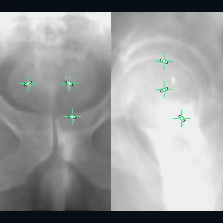
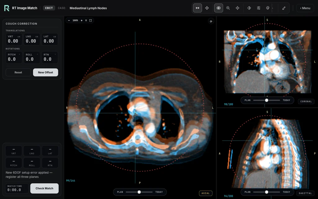
The landing page includes unaltered screenshots of the actual trainer and instructor interface, captured on October 9, 2026. Femur 2D/2D images use the Soft-tissue-Sarcoma collection; Mediastinal Nodes images use the Mediastinal Lymph Node collection; Prostate Full Treatment and Machine Room use Prostate Anatomical Edge Cases. The collection citations and licenses appear below. These views are confined to the thigh, thorax and pelvis.
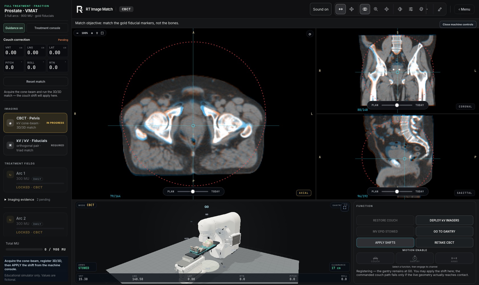
Captures use the existing isolated local fixture with fictional setup values. The Full Treatment image uses an acquisition-timing shortcut and depicts matching in progress, not delivered treatment. Classroom names, plans, attendance and results are entirely fictional. The active Live Lab screenshot uses the same reviewed Mediastinal Nodes thoracic images cited above. Native screenshots and element crops are used without generated anatomy or UI. The October 9 Plan and Today pair is retained below as historical media. The earlier pair used a display crop to remove its photographed toolbar and slider. Those registered screenshot files remain unchanged. The training equipment is representative geometry, independent of equipment manufacturers. Exact source revisions, capture timestamps and file hashes are recorded in docs/landing-cinematic-captures.json and docs/data-rights-register.json.
Motion uses GSAP and ScrollTrigger under the GSAP Standard License and Lenis under MIT. Earlier landing designs adapted David Haz's React Bits TiltedCard and SpotlightCard; those decorative effects have been removed, and their attribution is retained under MIT + Commons Clause. The Higgsfield storyboard contains the design plan; it does not generate or alter the trainer imagery. See software notices and assets/vendor/cinematic-provenance.json for exact source provenance.
- cinematic-planar-20261009.jpg
- cinematic-planar-detail-20261009.jpg
- cinematic-cbct-20261009.jpg
- cinematic-cbct-detail-20261009.jpg
- cinematic-plan-20261009.jpg
- cinematic-today-20261009.jpg
- cinematic-treatment-20261009.jpg
- cinematic-machine-20261009.jpg
- cinematic-machine-detail-20261009.jpg
- cinematic-machine-inside-20261009.jpg
- cinematic-classroom-plan-20261009.webp
- cinematic-classroom-active-20261009.jpg
- cinematic-classroom-review-20261009.webp
Full Treatment: October 10 capture
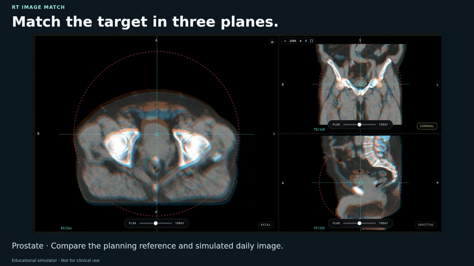
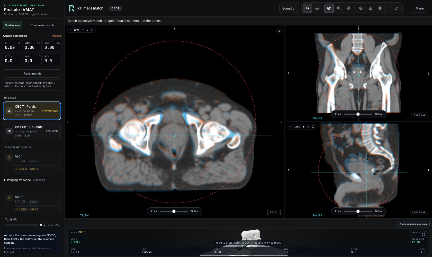
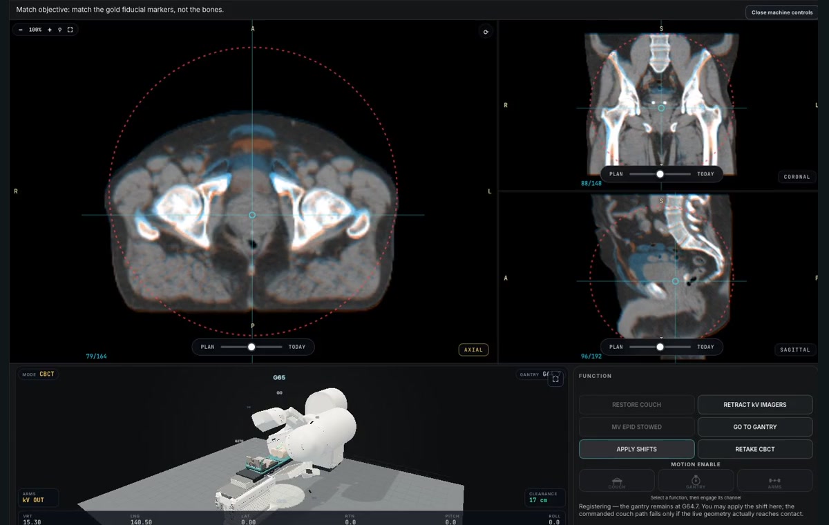
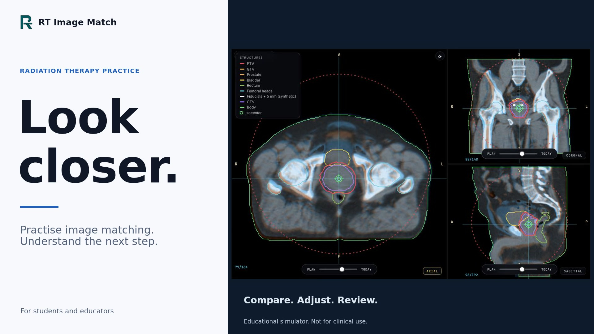
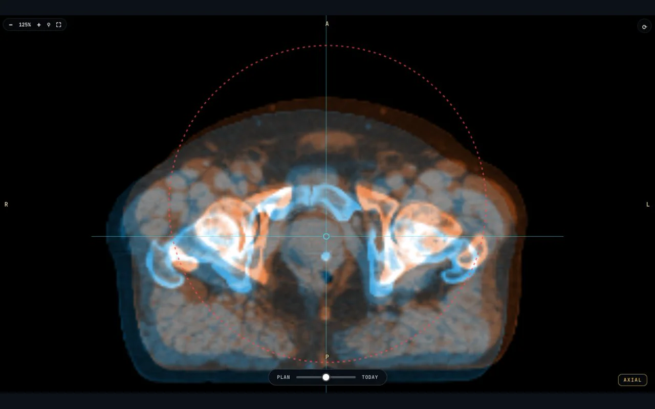
The landing page's Full Treatment image is a fresh native screenshot of the Prostate workflow. The existing machine controls are expanded and the Room camera is zoomed out with the trainer's own wheel control, so the 3D linac and couch appear beneath all three CBCT matching views. Source imaging is from Prostate Anatomical Edge Cases (TCIA), licensed CC BY 4.0; the full collection citation appears below. Plan and Today use the same pelvic source with a fictional setup error. The local fictional-account fixture shortens acquisition timing only: matching is in progress, couch correction is pending and no treatment is delivered. No generated imagery, montage, altered layout, source head or facial imaging, real account or learner record is included. The machine remains representative teaching geometry. Capture state, source revision, model and image hashes are recorded in docs/landing-treatment-capture.json.
Femur and Full Treatment: guided scroll recordings
The October 12 guided stories use new native trainer recordings from an isolated fictional-account fixture. Femur shows a scripted translation and rotation followed by its actual accepted registration review. Prostate shows native acquisition, registration, held couch correction, fiducial verification and simulated delivery; machine actions use the trainer’s full-screen Room camera. The source collections and licences are listed below.
Desktop and phone versions use uniform crops and scaling, still-frame inspection holds and short crossfades. Phone views place simultaneous AP/lateral or three-plane source views together for readability. Playback timing is condensed; no anatomy, registration outcome or interface was generated or retouched. Perspective motion is presentation only. Full original screenshots remain available through Enlarge image. Source hashes, native outcome records, crop filters and timing are documented in docs/landing-guided-captures.json.
Guided story recordings and focused views
- Femur Desktop recording
- Femur Inspect Desktop still
- Femur Correct Desktop still
- Femur Review Desktop still
- Femur Mobile recording
- Femur Inspect Mobile still
- Femur Correct Mobile still
- Femur Review Mobile still
- Treatment Desktop recording
- Treatment Acquire Desktop still
- Treatment Match Desktop still
- Treatment Correct Desktop still
- Treatment Verify Desktop still
- Treatment Deliver Desktop still
- Treatment Mobile recording
- Treatment Acquire Mobile still
- Treatment Match Mobile still
- Treatment Correct Mobile still
- Treatment Verify Mobile still
- Treatment Deliver Mobile still
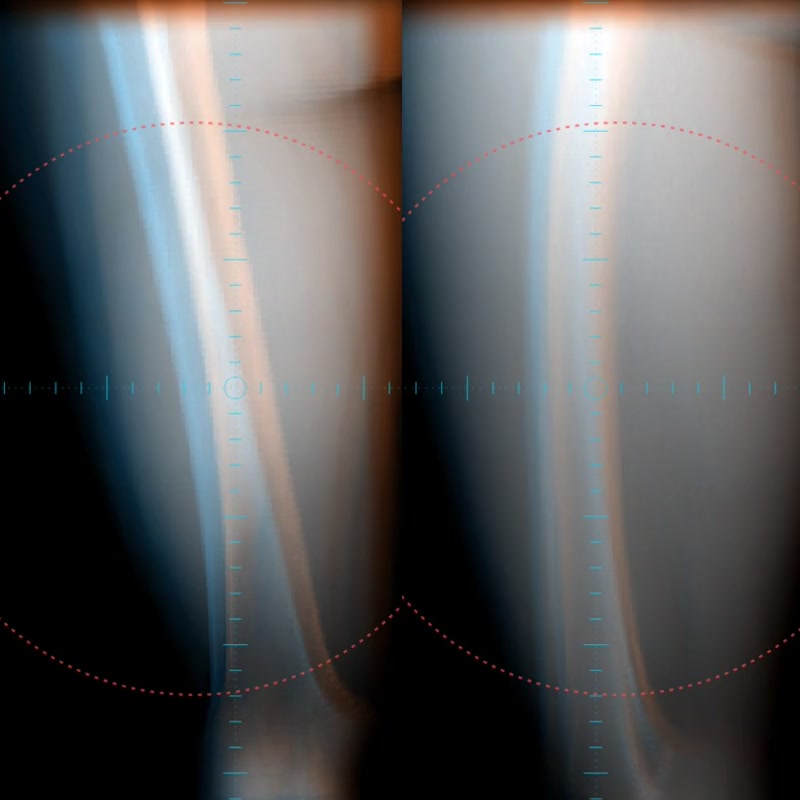
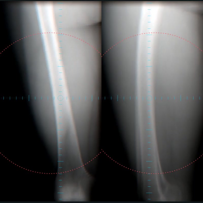
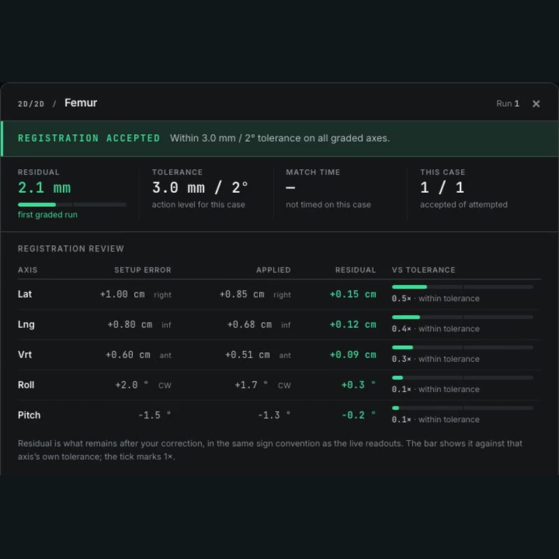
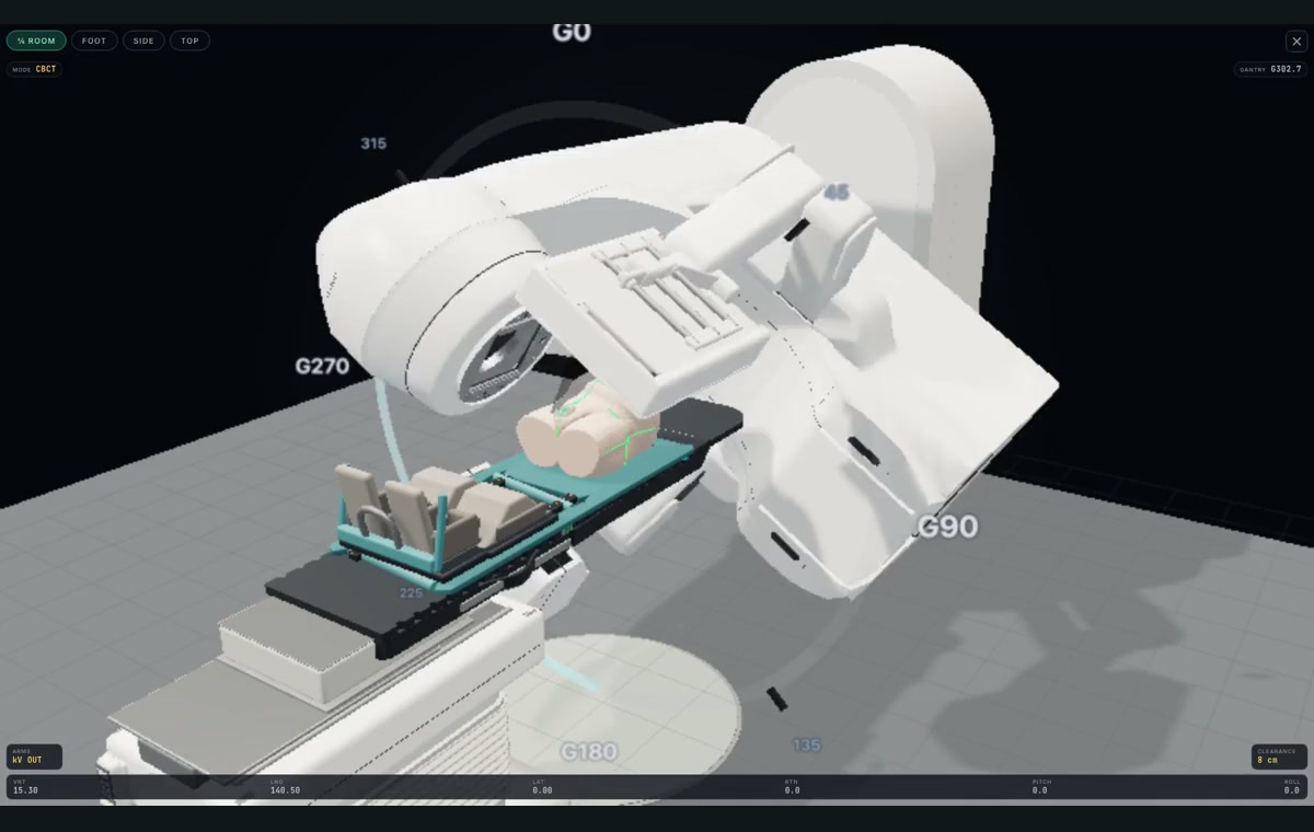
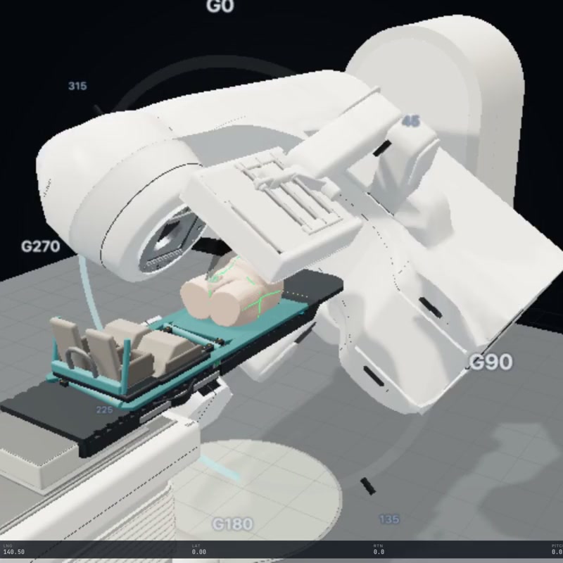
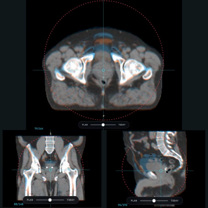
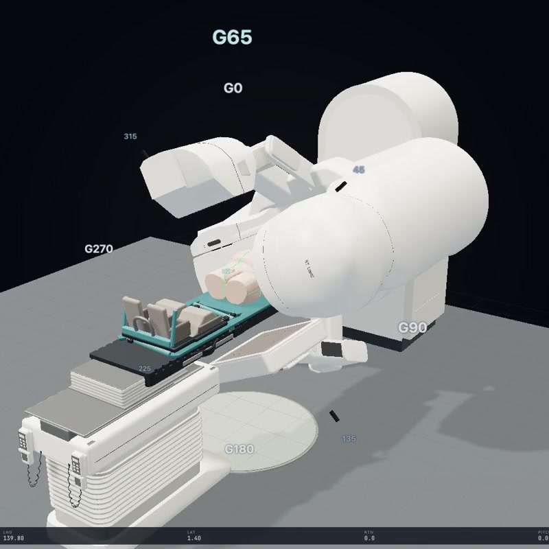
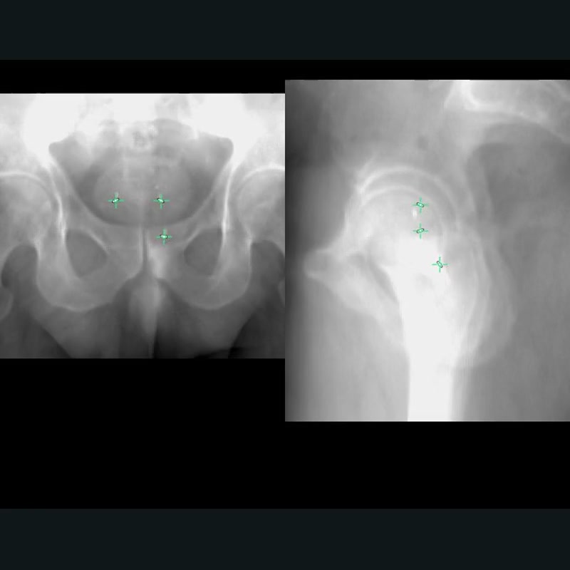
Worked example and Full Treatment: October 12 captures
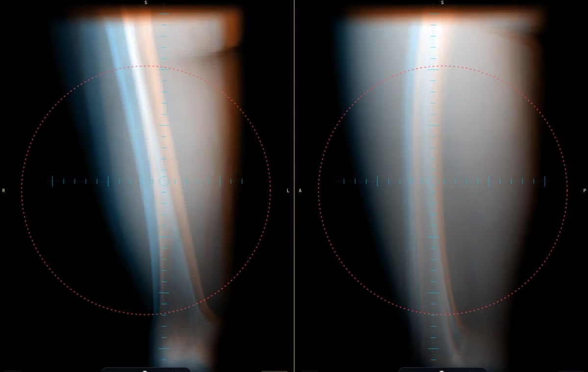
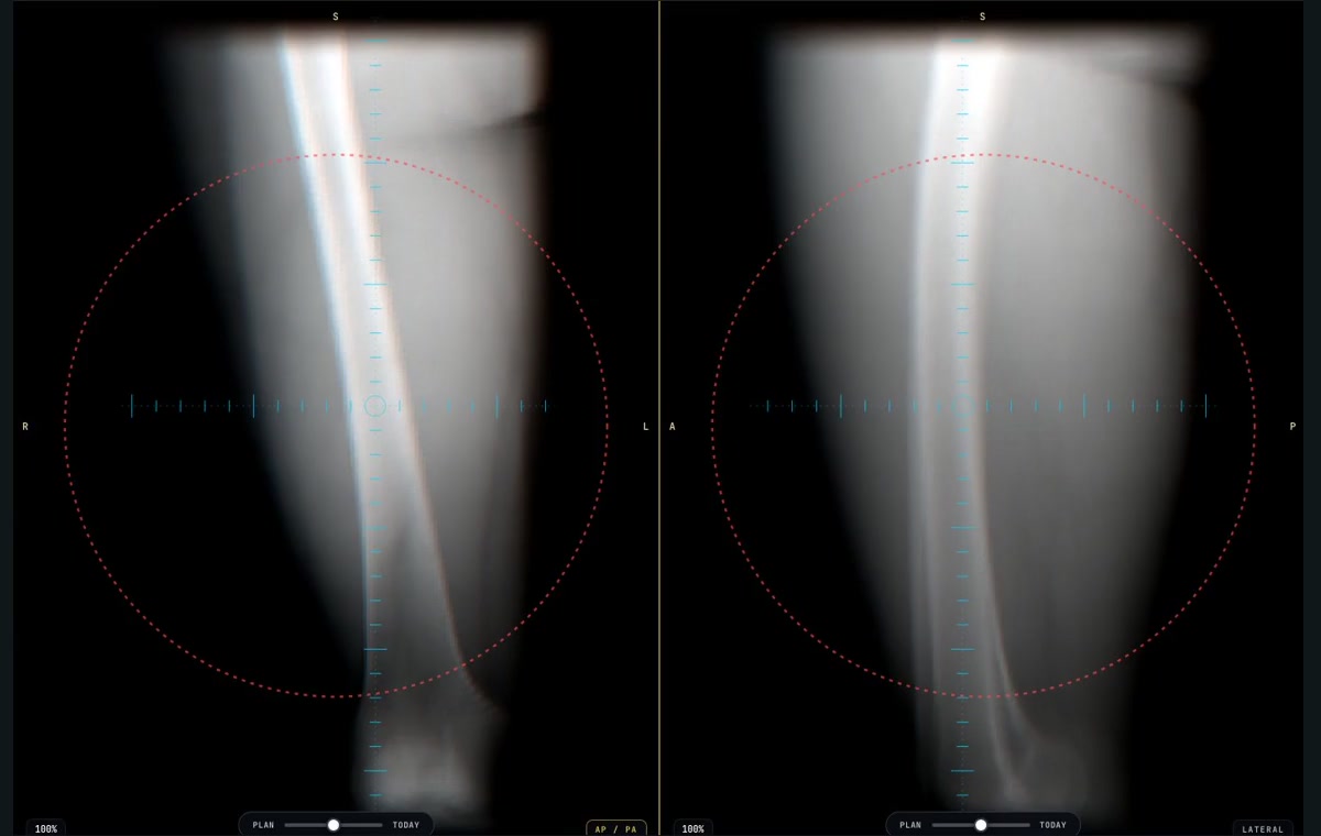
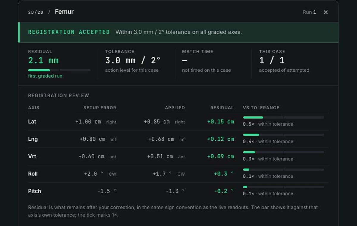
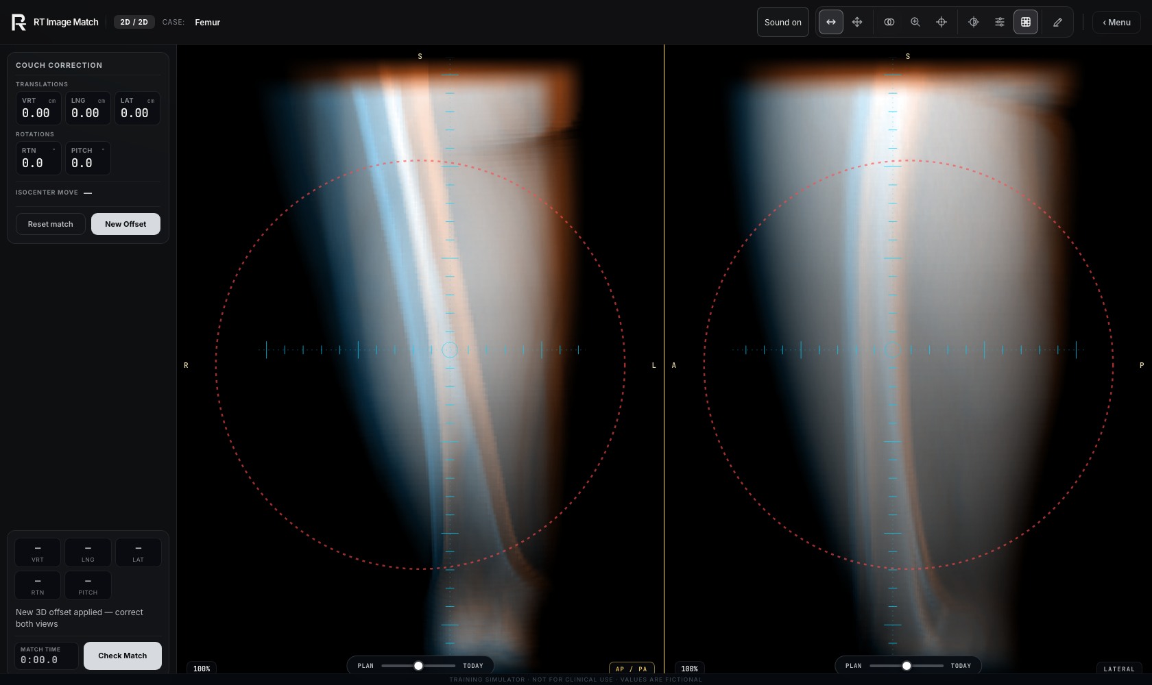
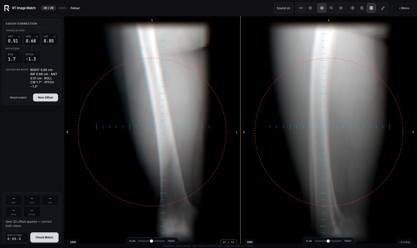
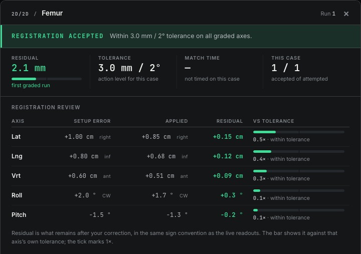
The Femur example shows the native 2D/2D trainer before and after a scripted correction, followed by its actual registration verdict and residual table. The review is a crop of that native report. The fictional result belongs only to this recorded example. Femur imaging is adapted from the Soft-tissue-Sarcoma collection, licensed CC BY 3.0; the full citation and source conditions appear below.
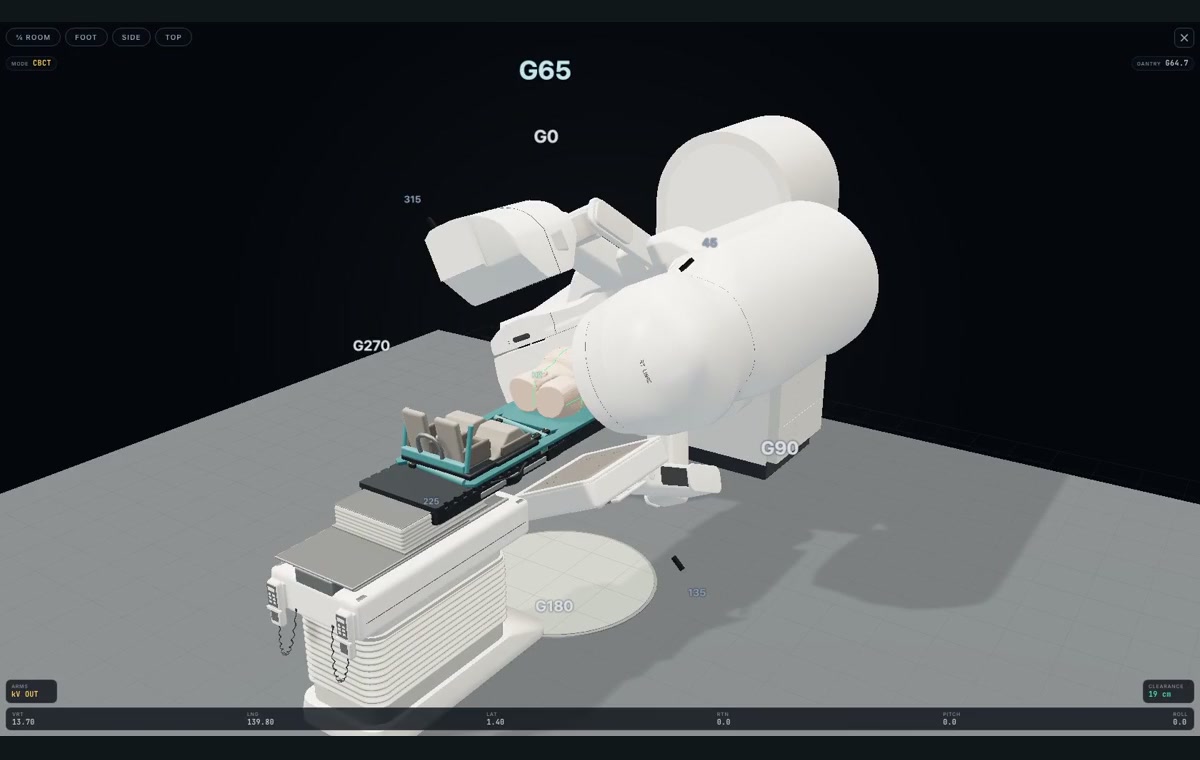
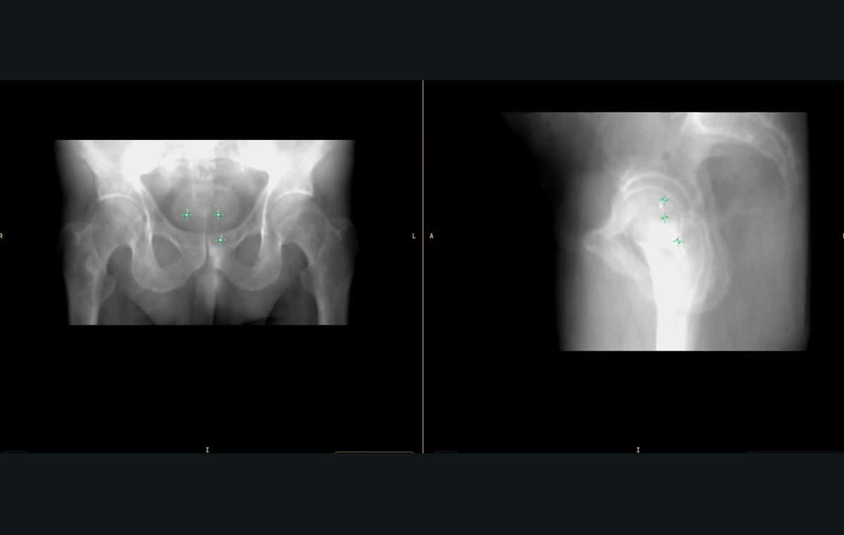
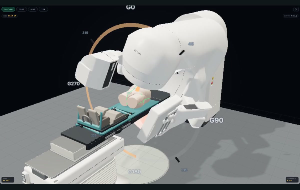
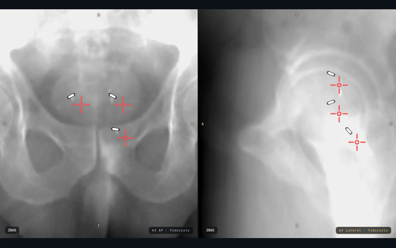
The five Prostate stages show actual CBCT acquisition, image matching, held couch correction, post-correction kV fiducial verification and simulated VMAT delivery. Source imaging is adapted from Prostate Anatomical Edge Cases, licensed CC BY 4.0, with the full citation below. Recordings use deterministic fictional setup values and scripted controls in the isolated local account fixture. Acquisition waits and field preparation are condensed; native held-motion, clearance and delivery gates remain authoritative. Playback is time-condensed and silent. The camera uses the trainer’s own Room view and zoom controls.
These native screenshots and recordings contain thigh and pelvic source anatomy only, with no source head or facial field, production account or real learner record. No anatomy or interface was generated or retouched. The equipment is representative teaching geometry. This is a partial educational walkthrough, not a complete clinical protocol. Exact source and media hashes and the native outcome record are in docs/landing-practice-captures.json and the rights register. Earlier October 9 and 10 captures remain available for existing links.
- Femur: Inspect · Correct · Review
- Acquire: Still · Recording
- Match: Still · Recording
- Correct: Still · Recording
- Verify: Still · Recording
- Simulated delivery: Still · Recording
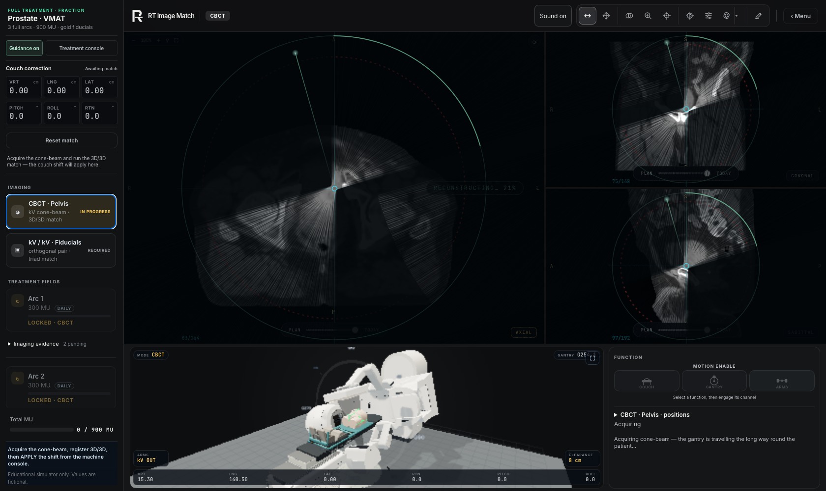
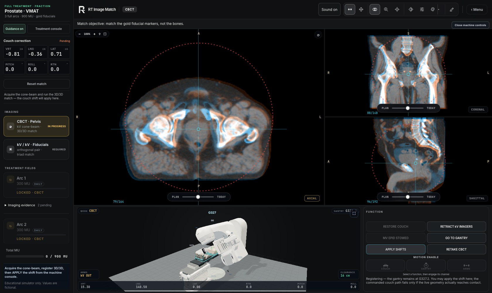
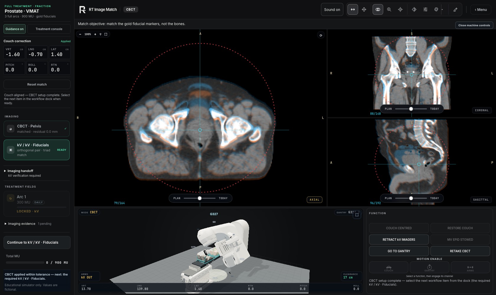
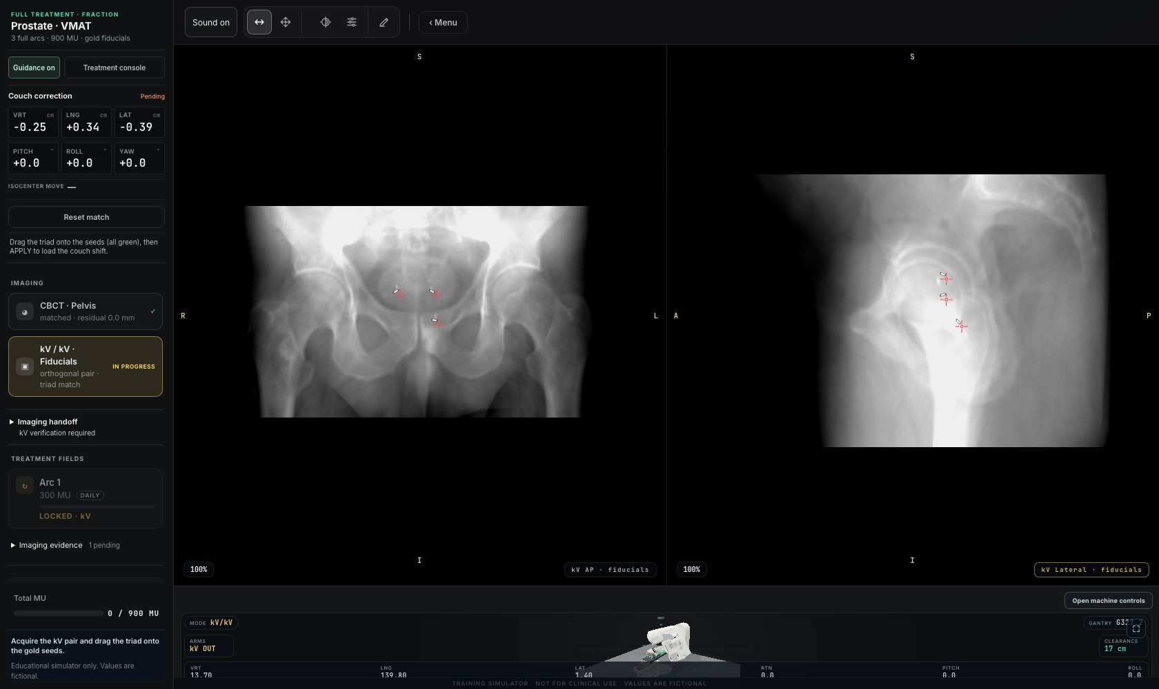
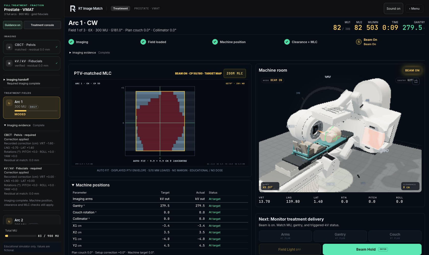
Machine Room: connected component tour
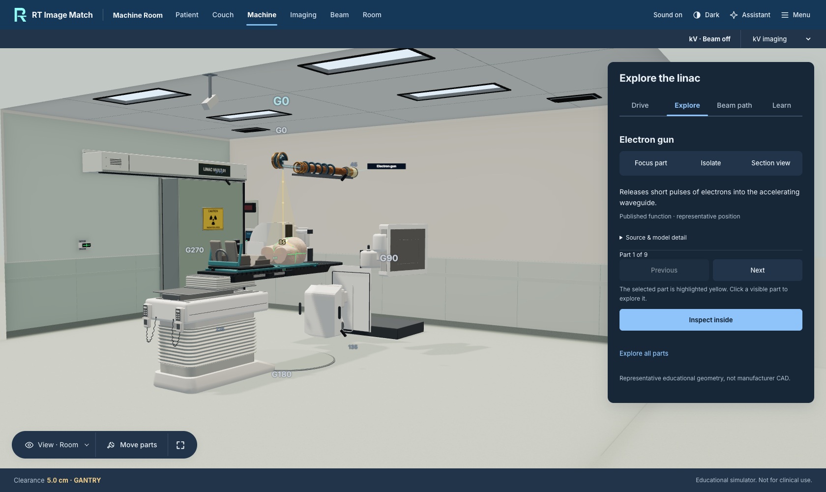
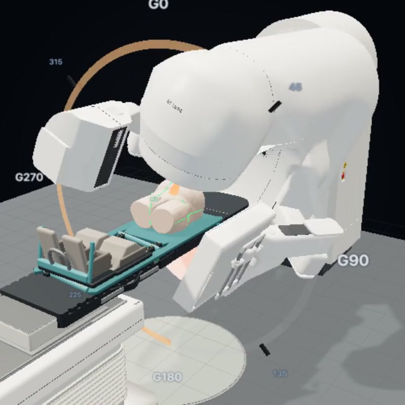
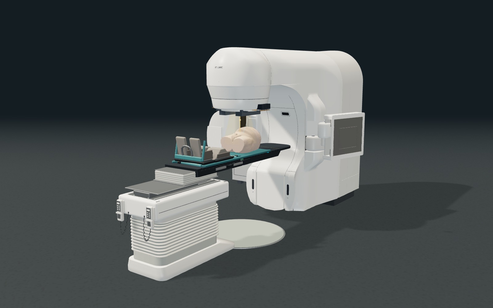
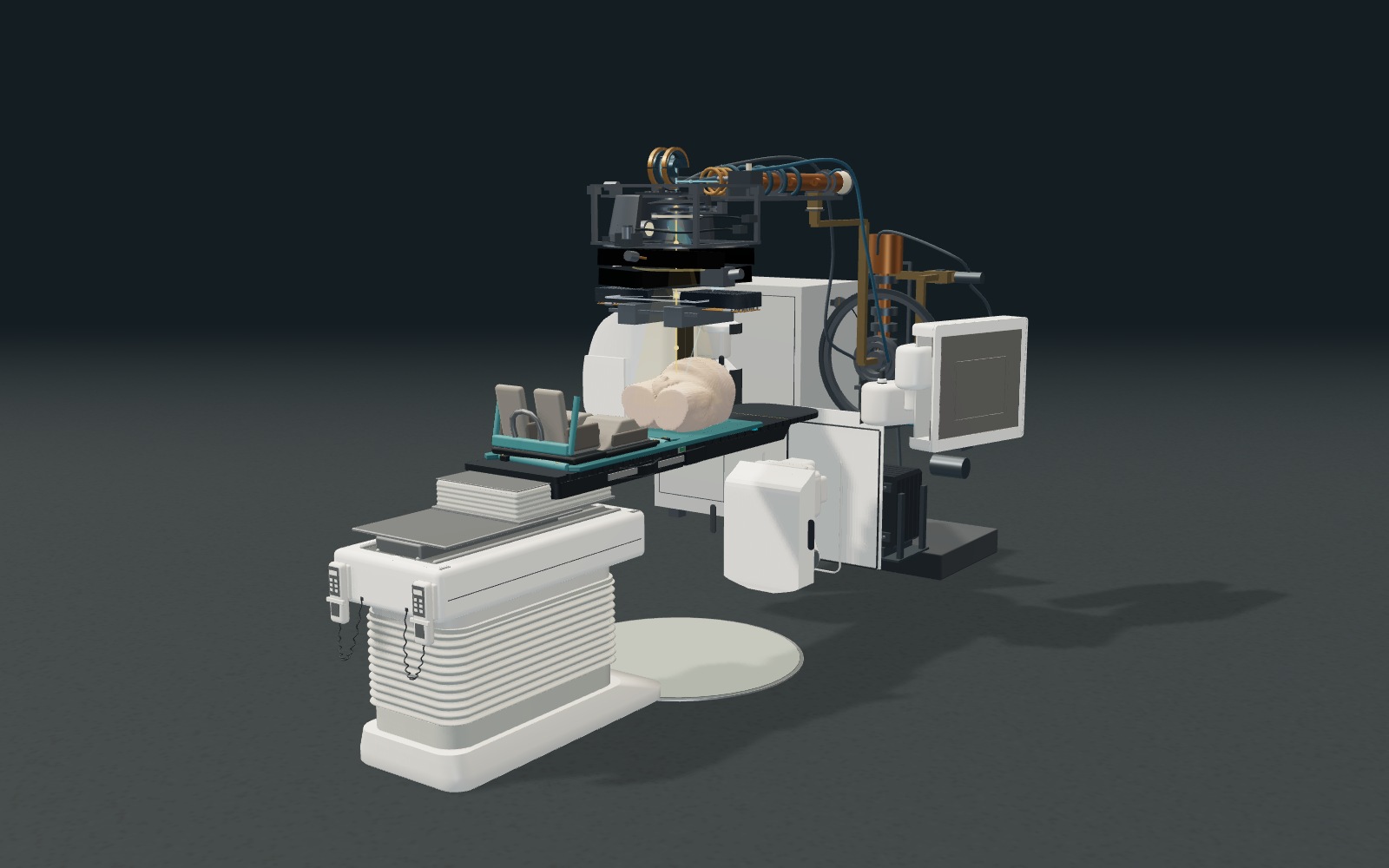
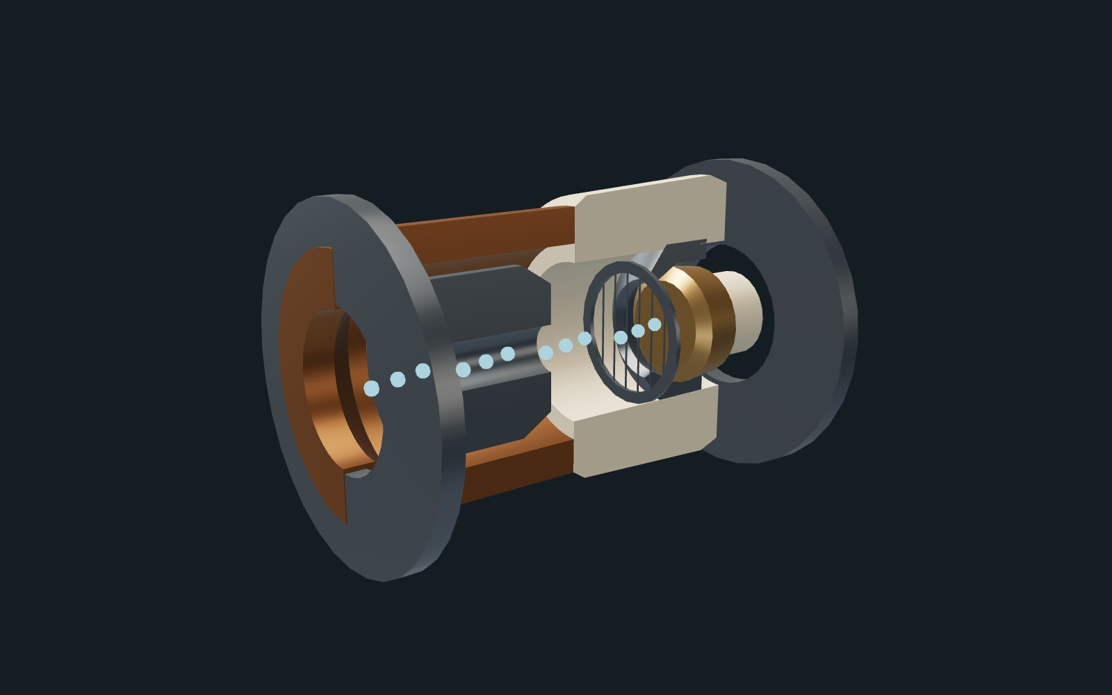
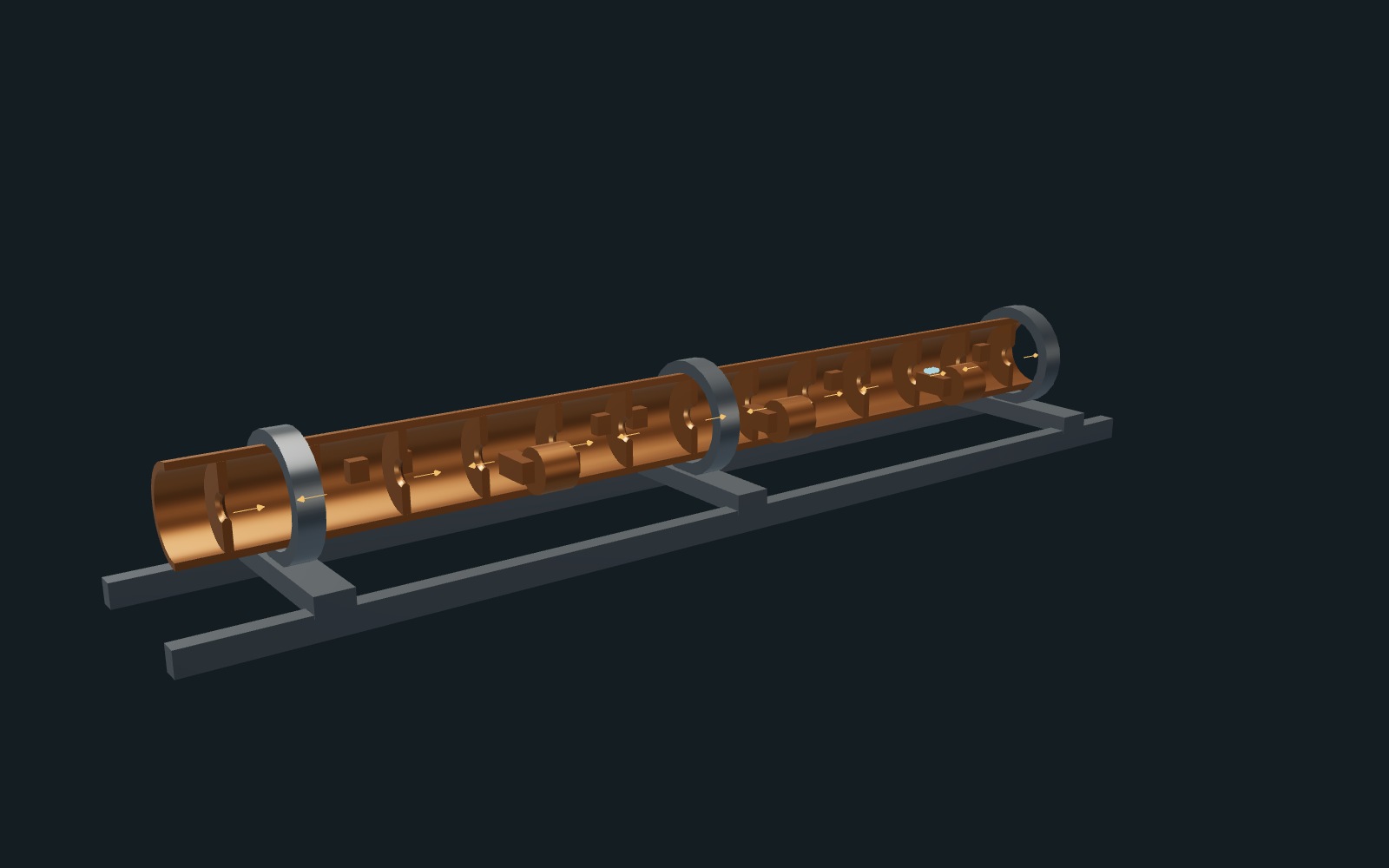
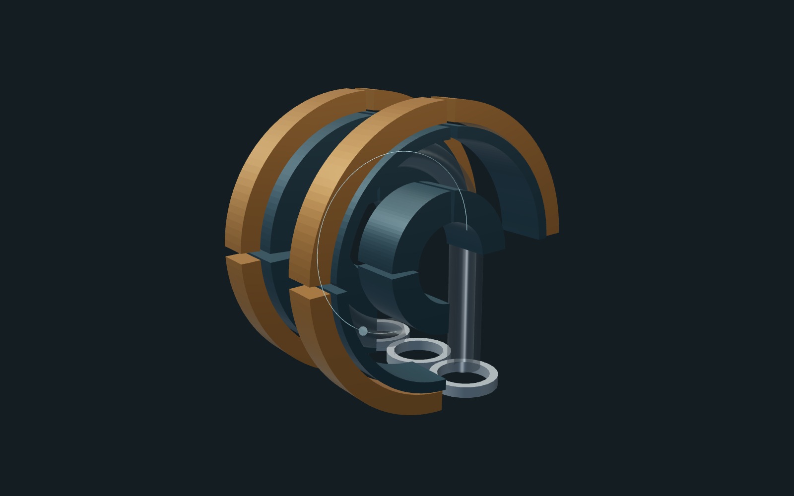
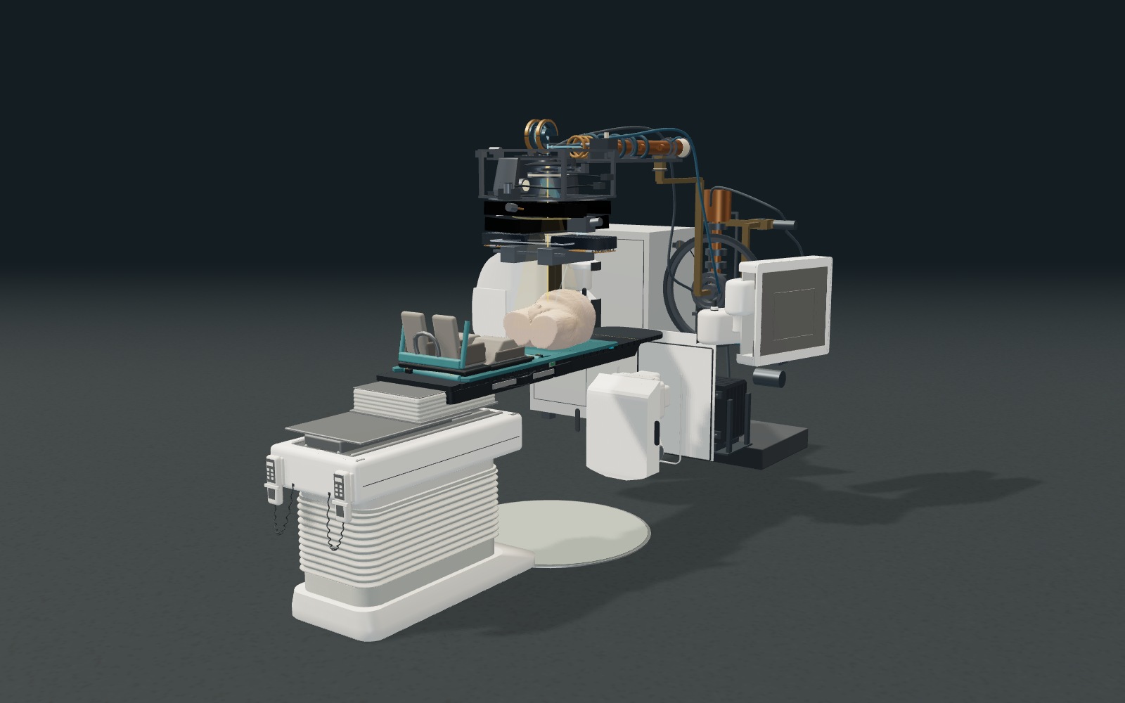
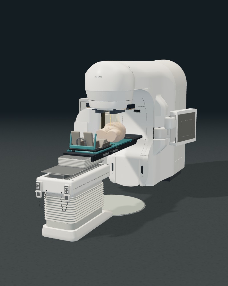
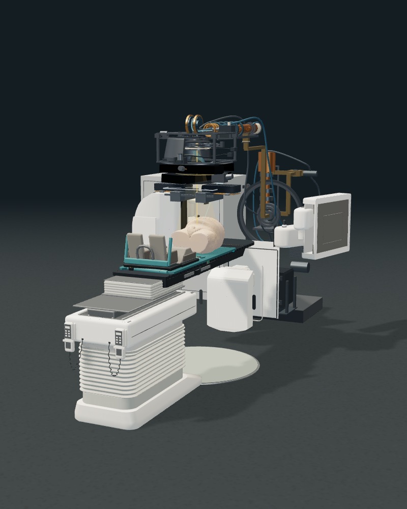
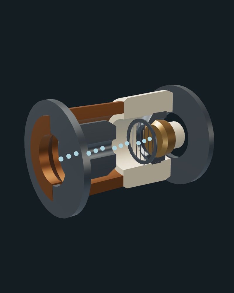
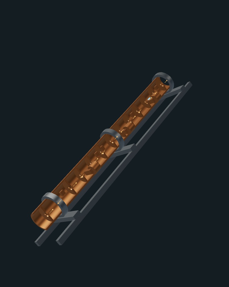
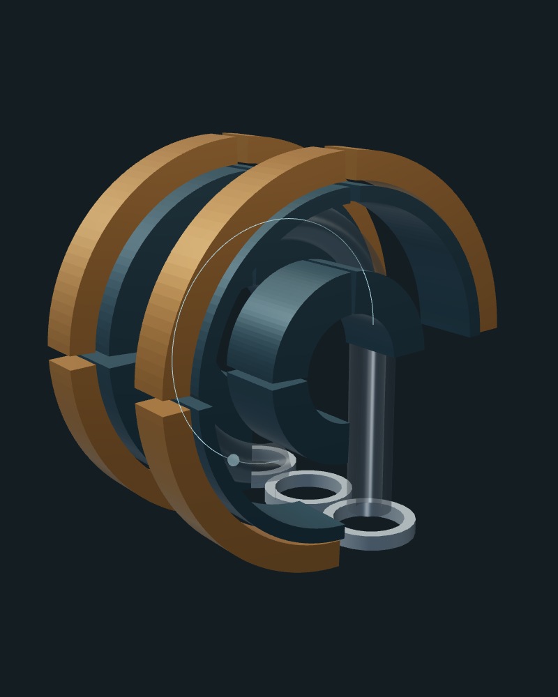
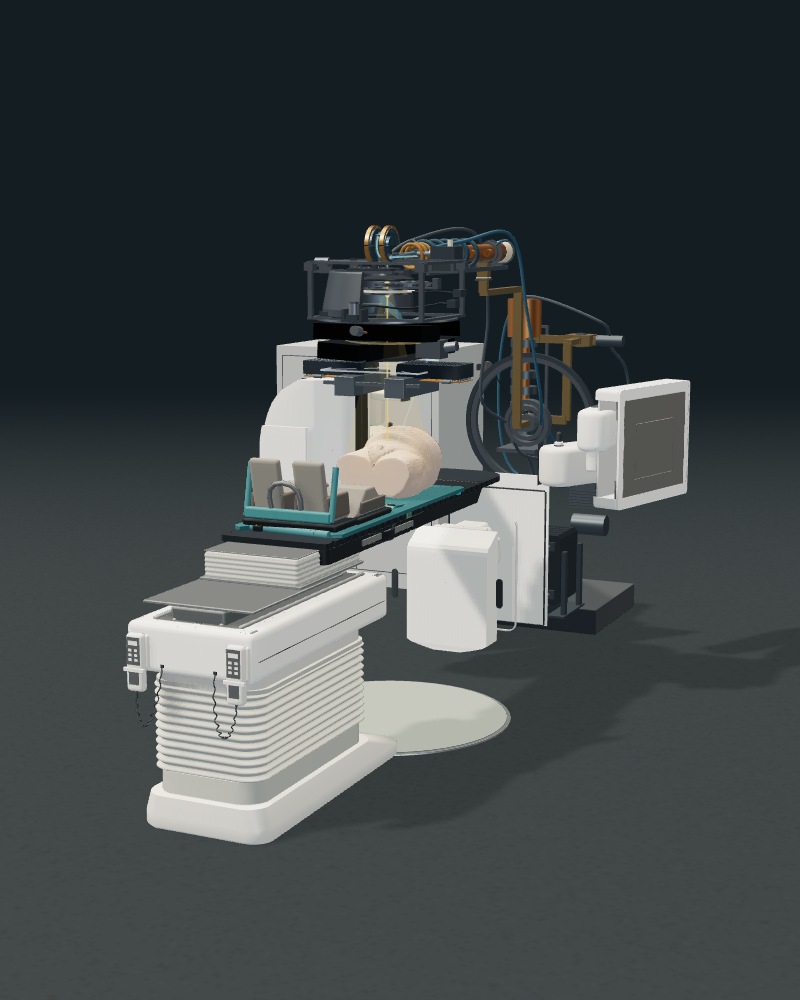
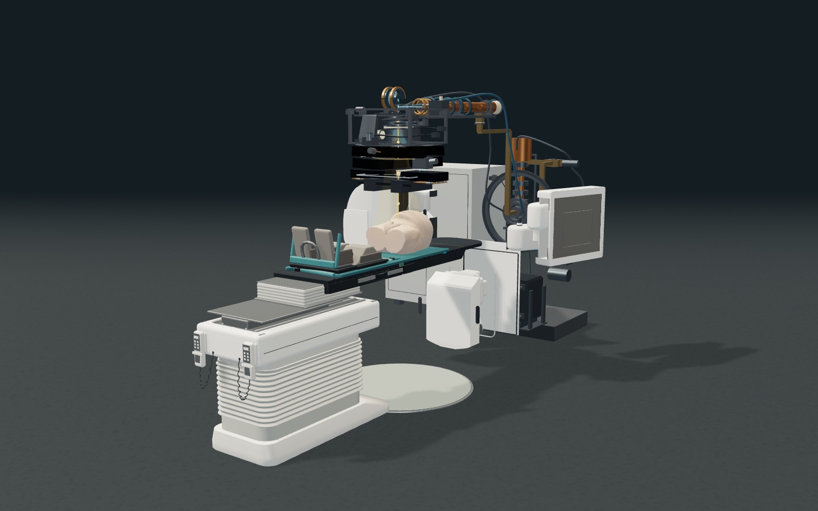
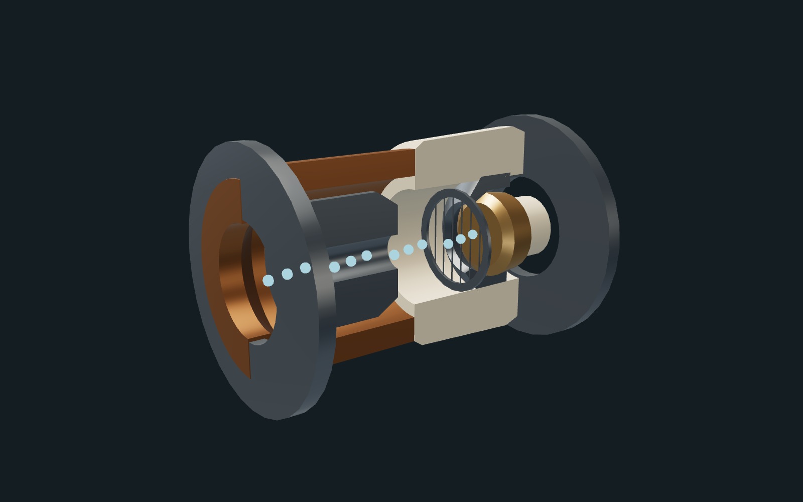
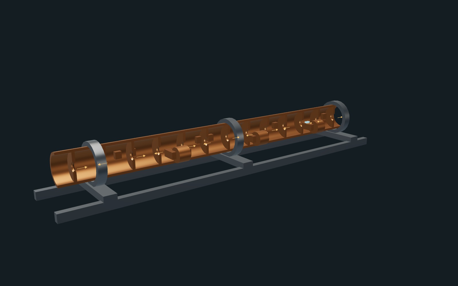
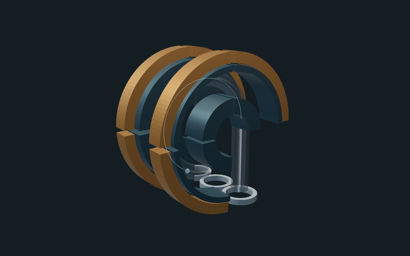
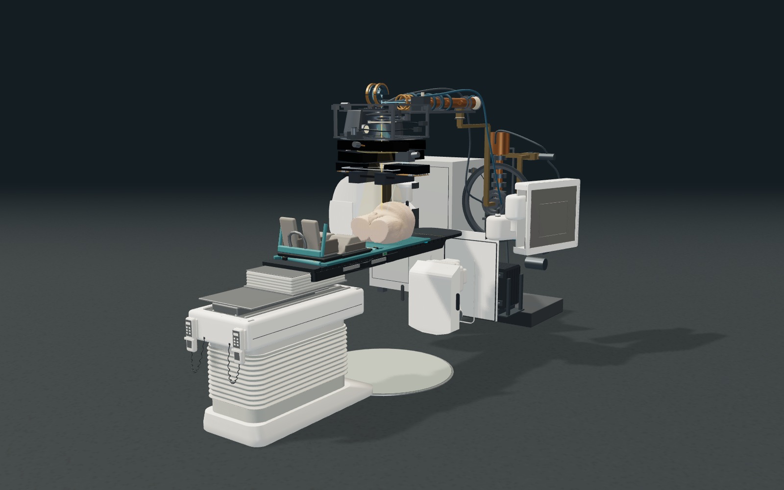
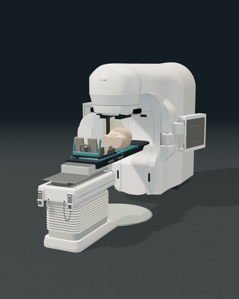
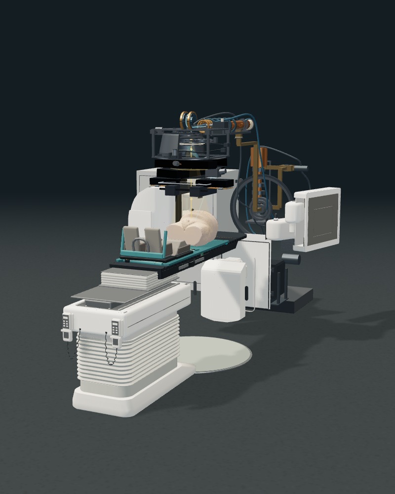
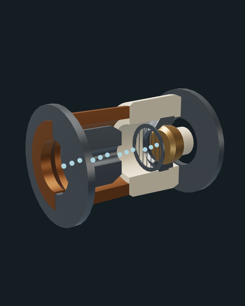
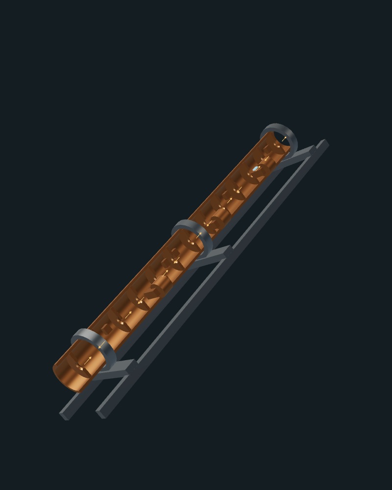
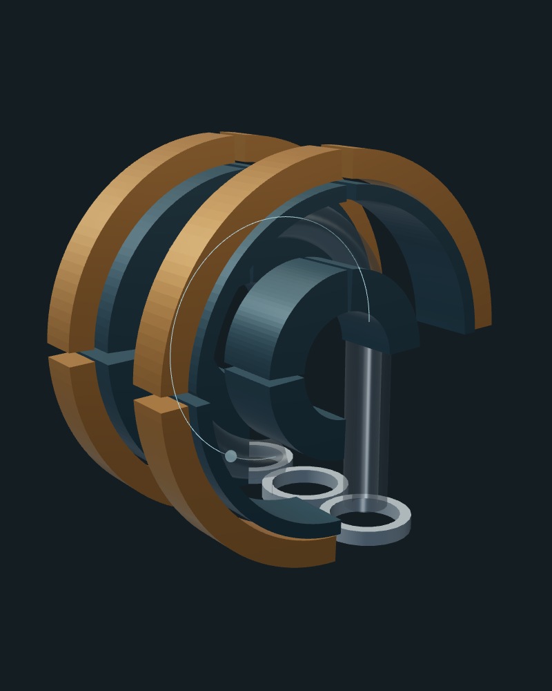
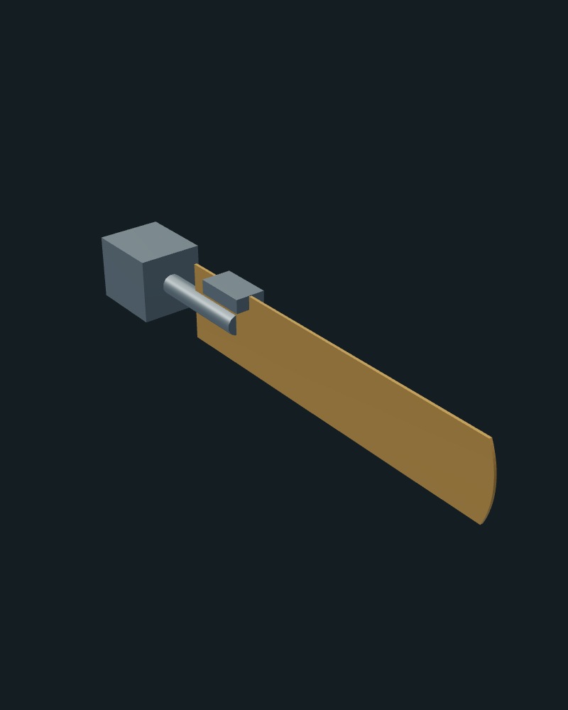
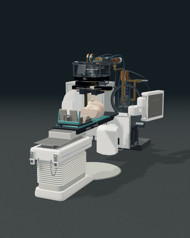
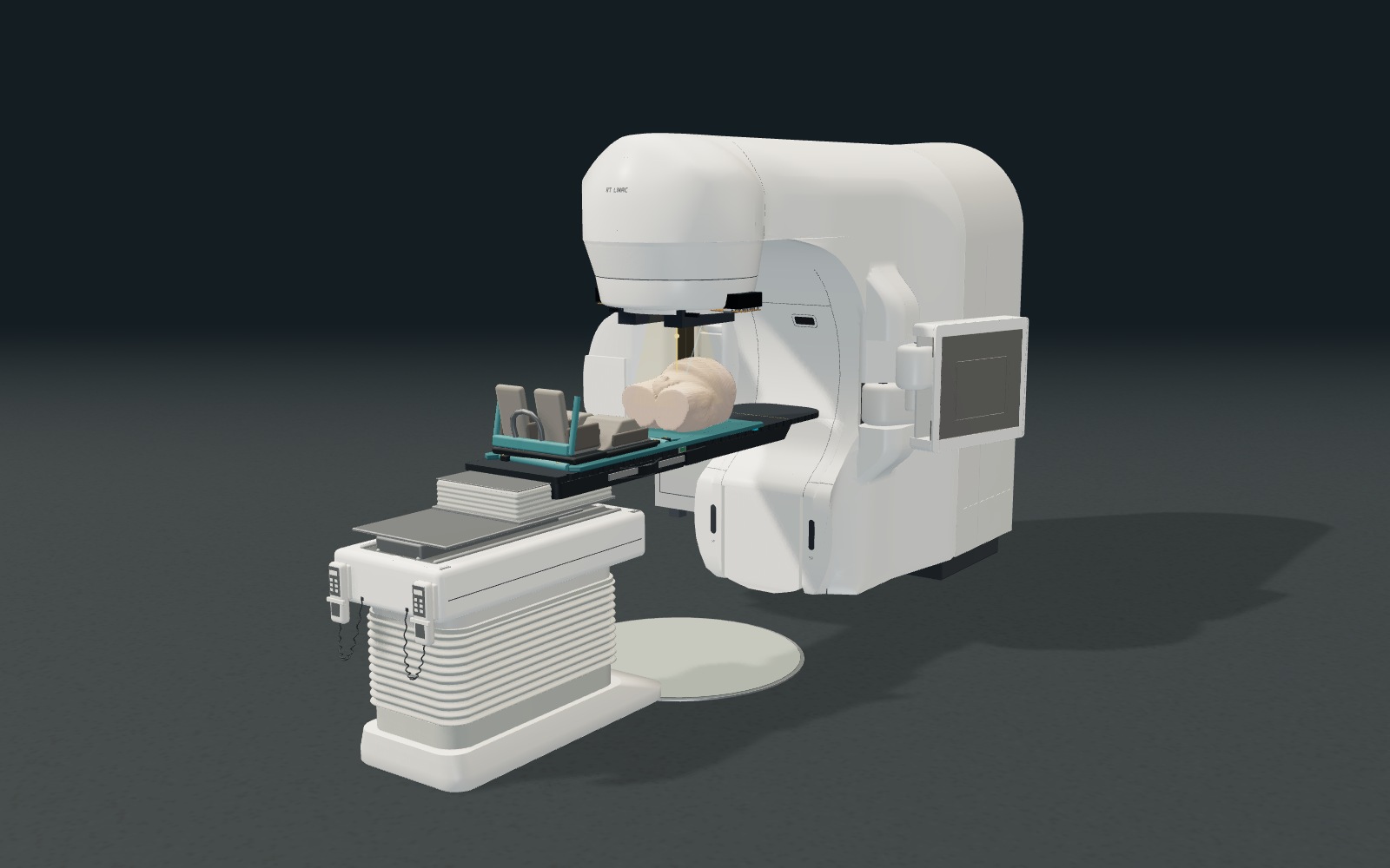
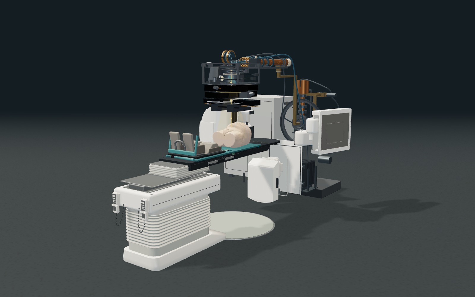
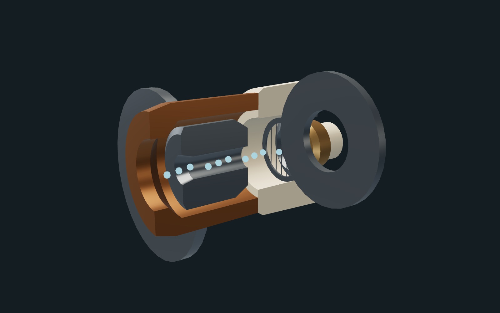
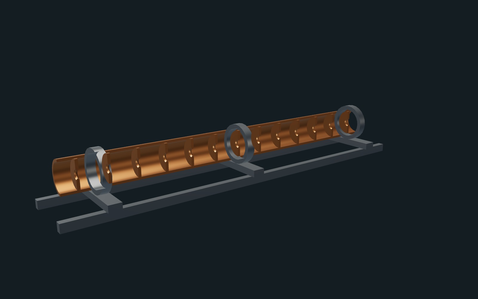
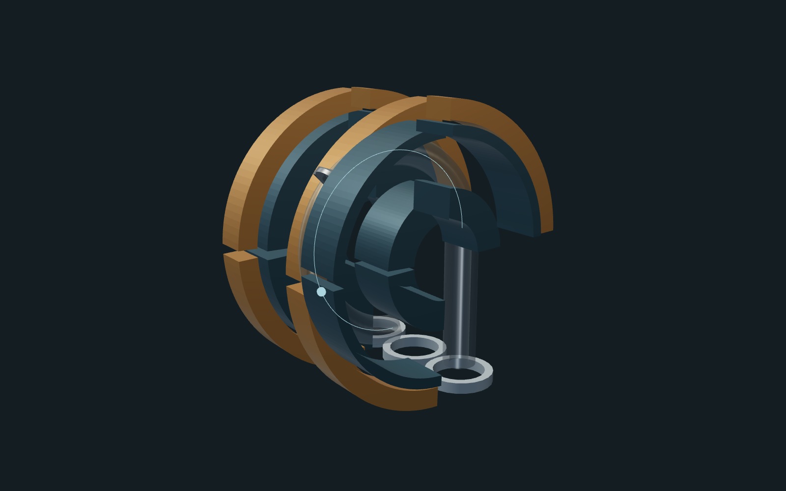
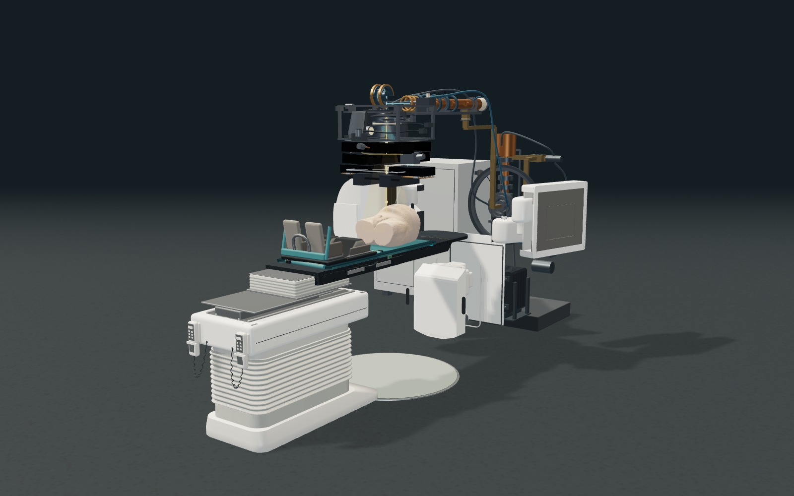
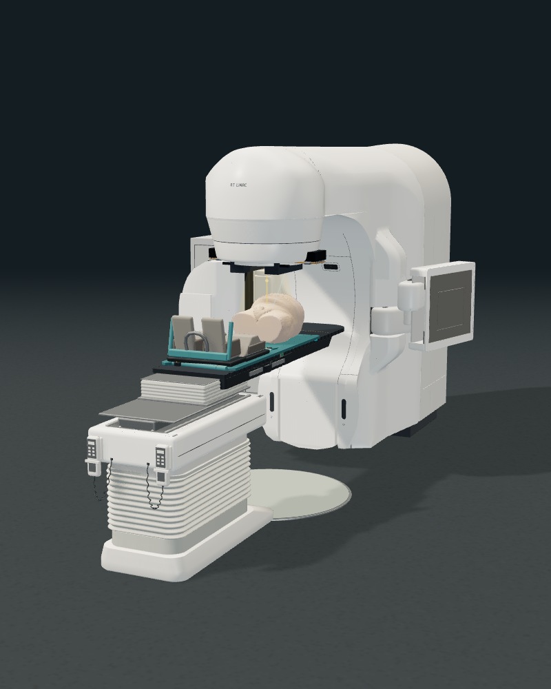
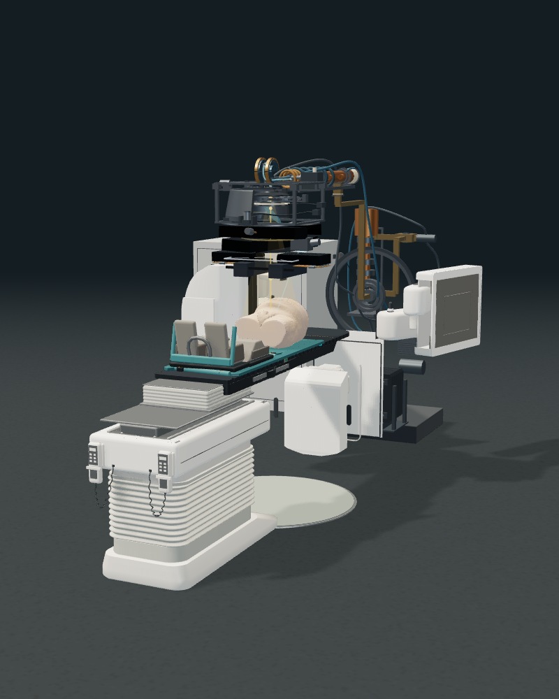
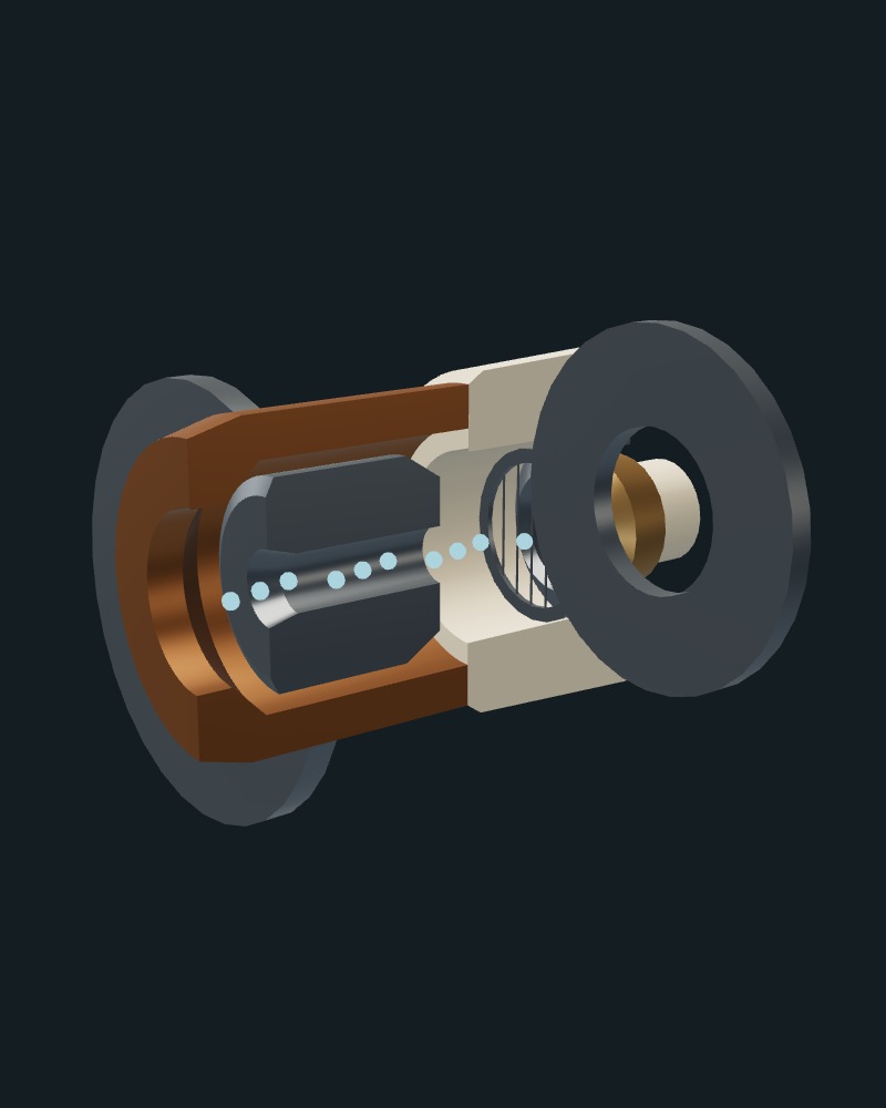
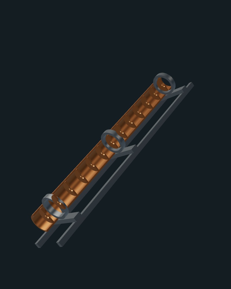
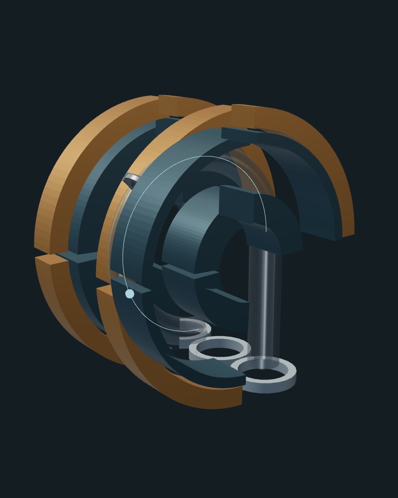
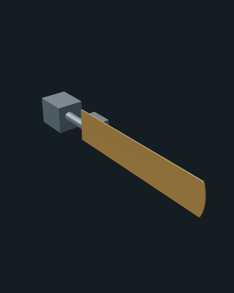
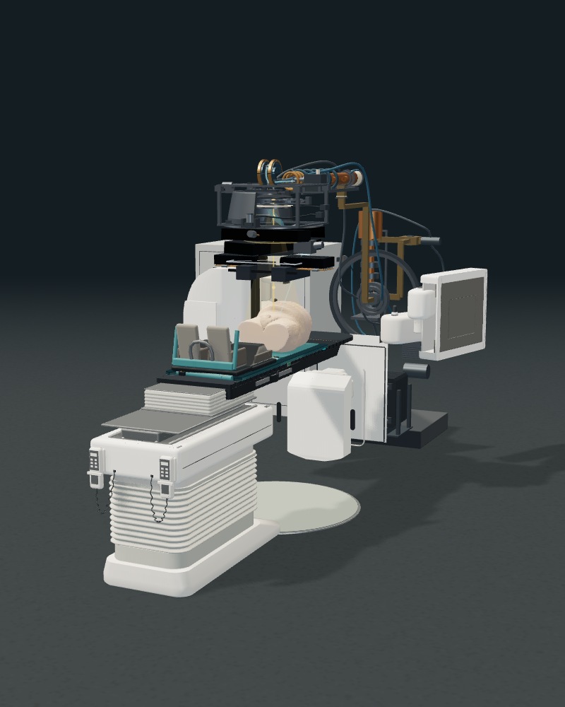
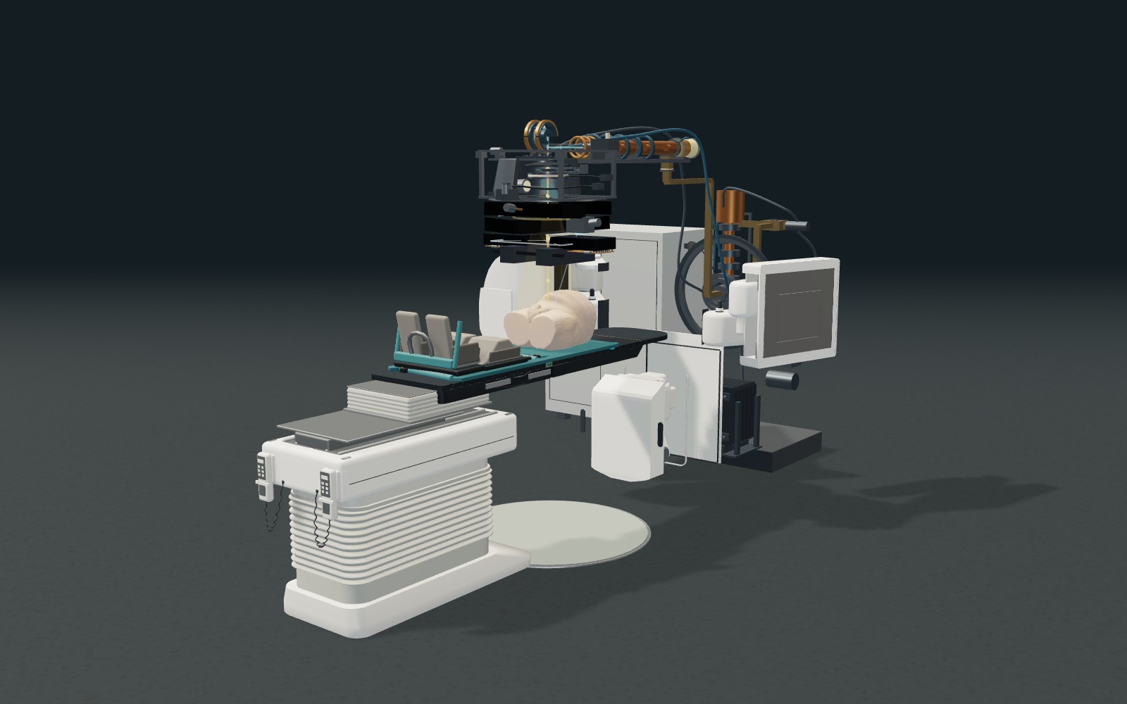
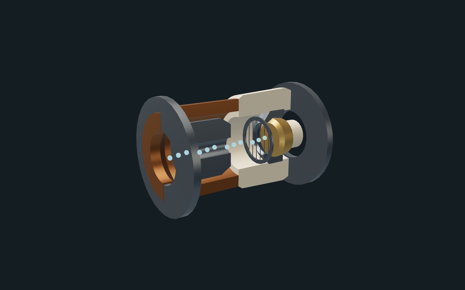
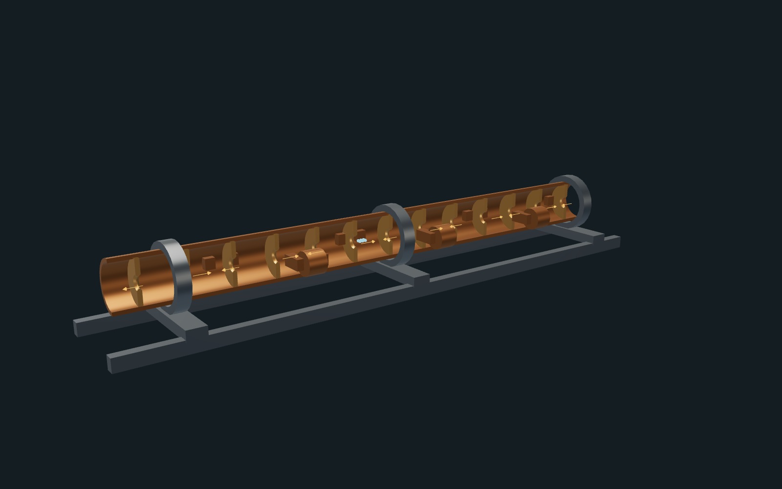
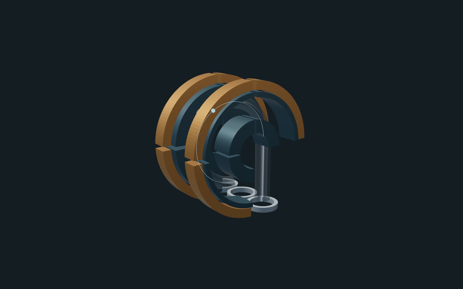
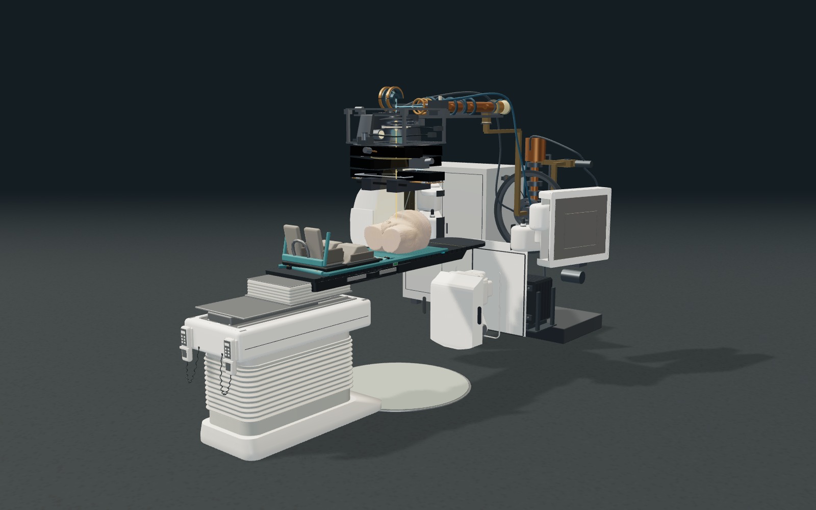
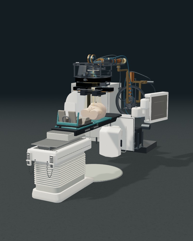
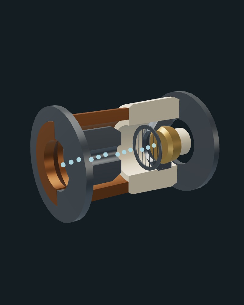
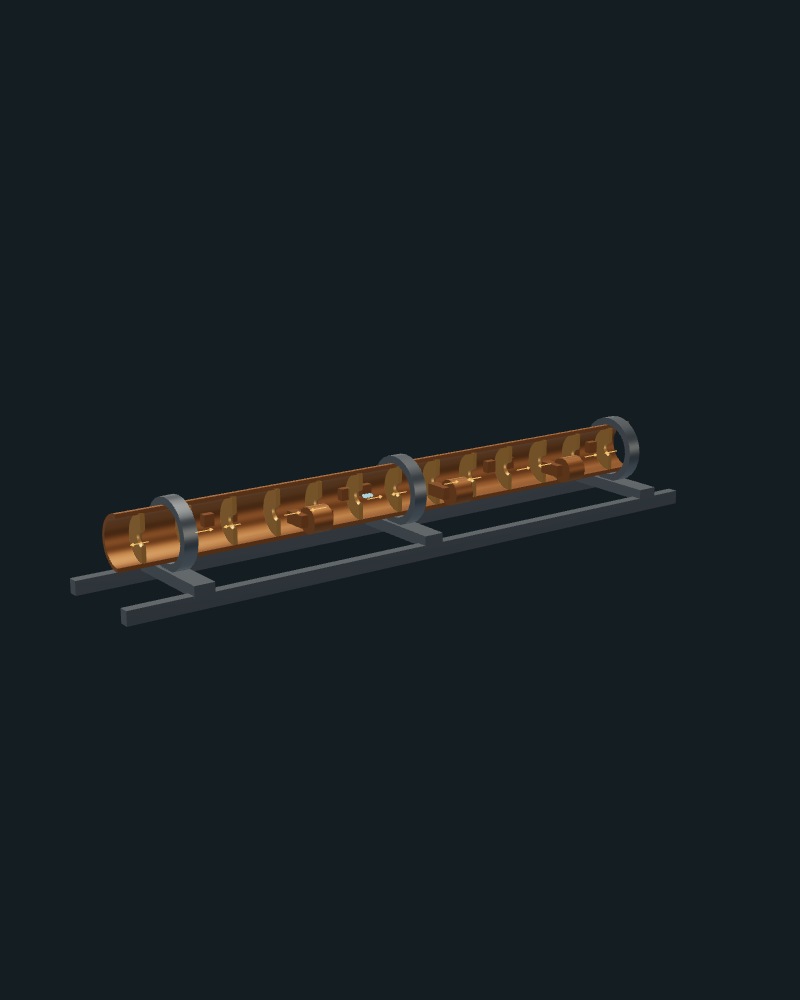
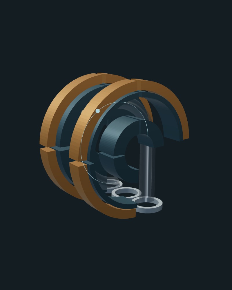
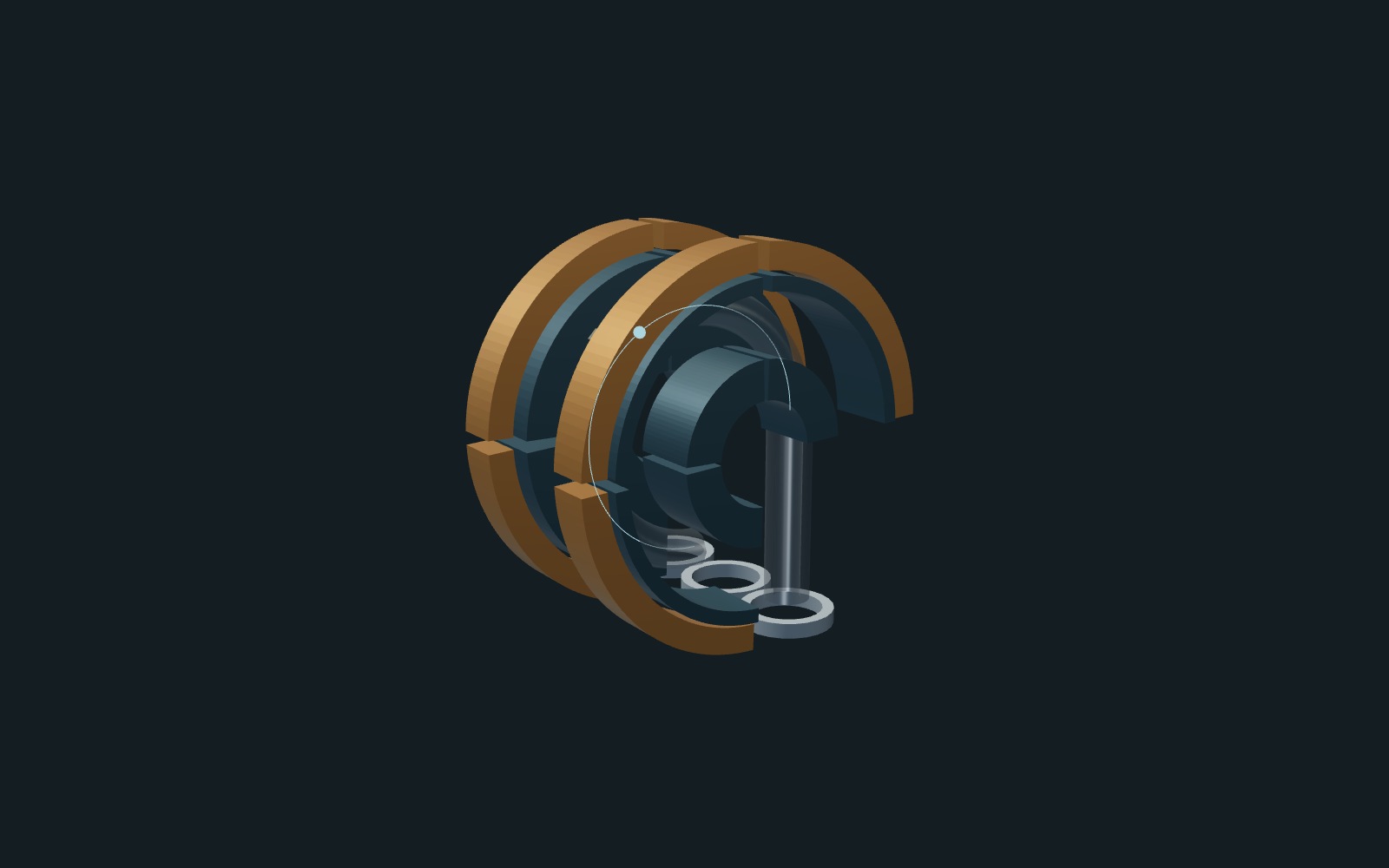
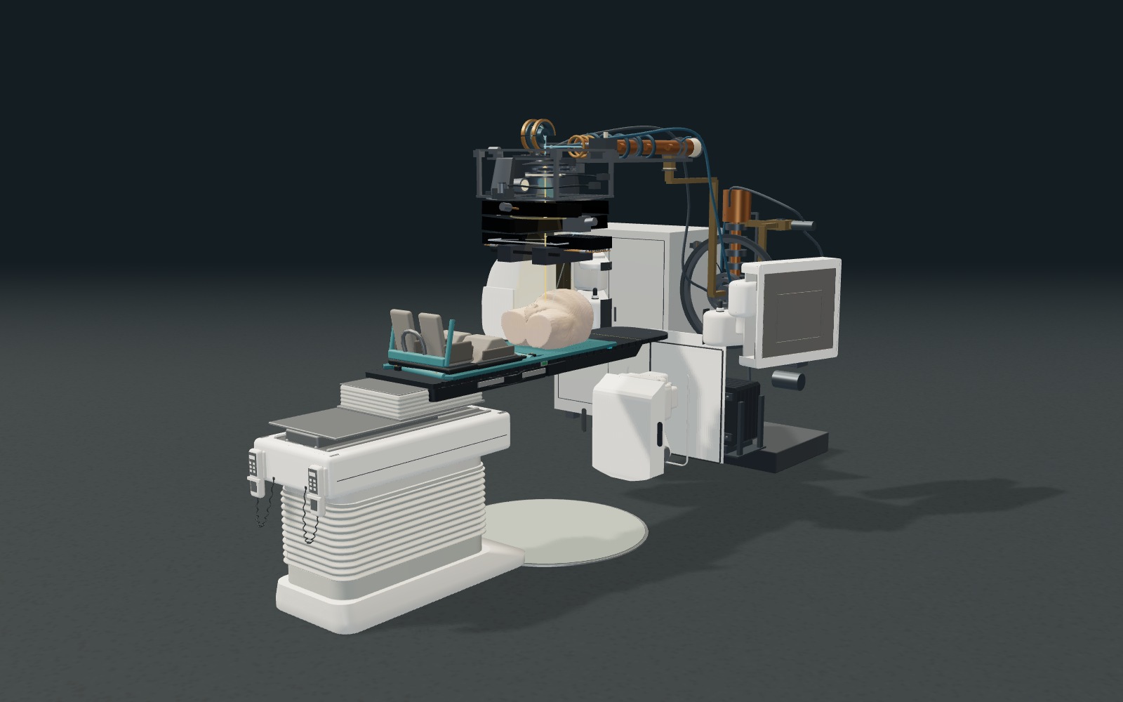
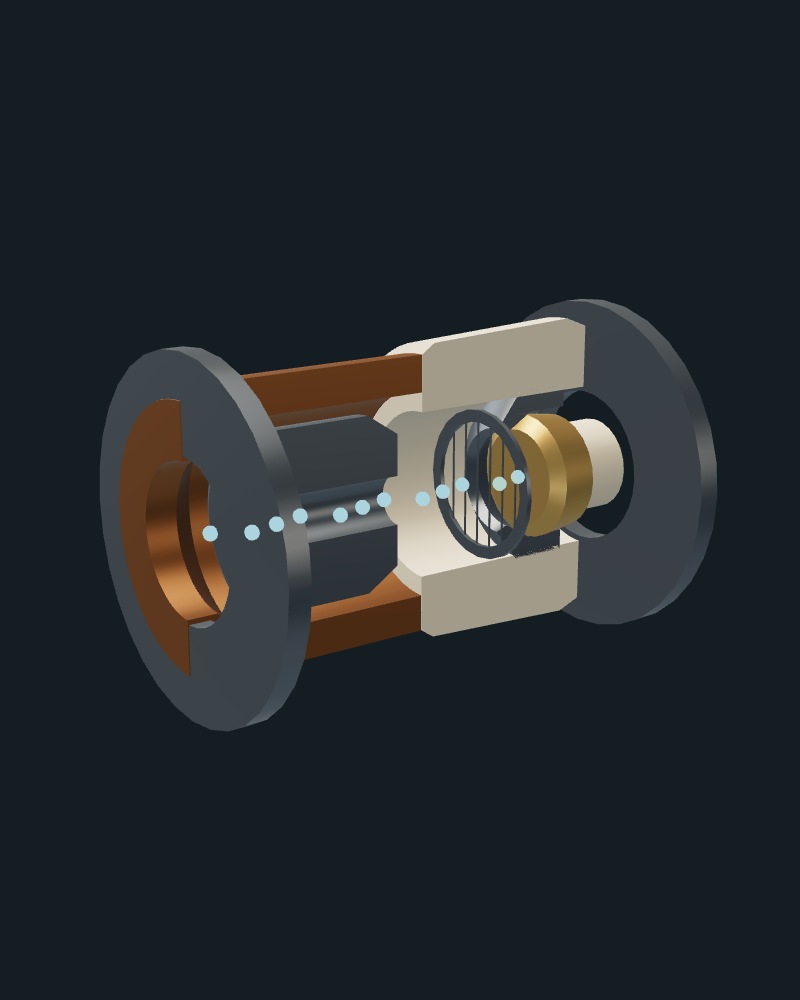
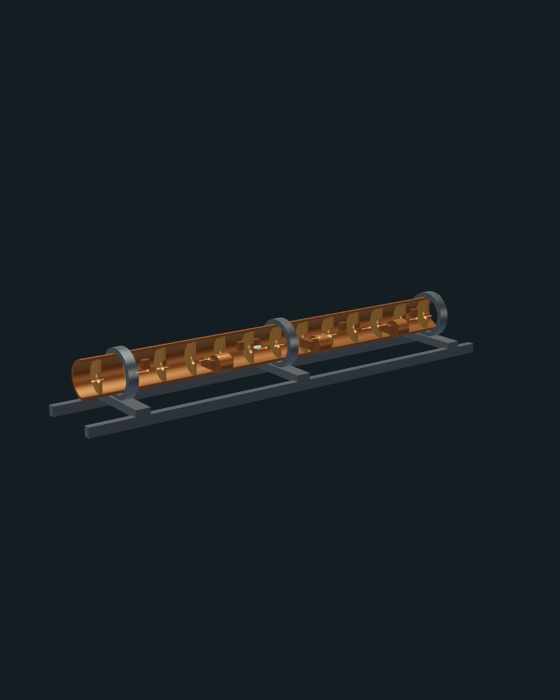
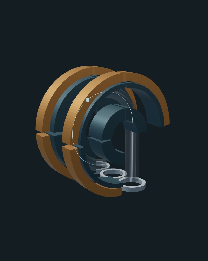
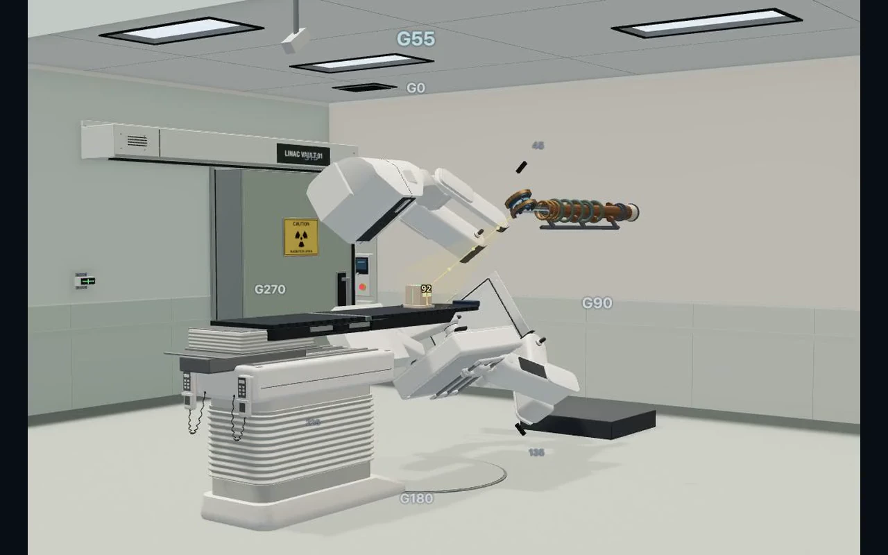
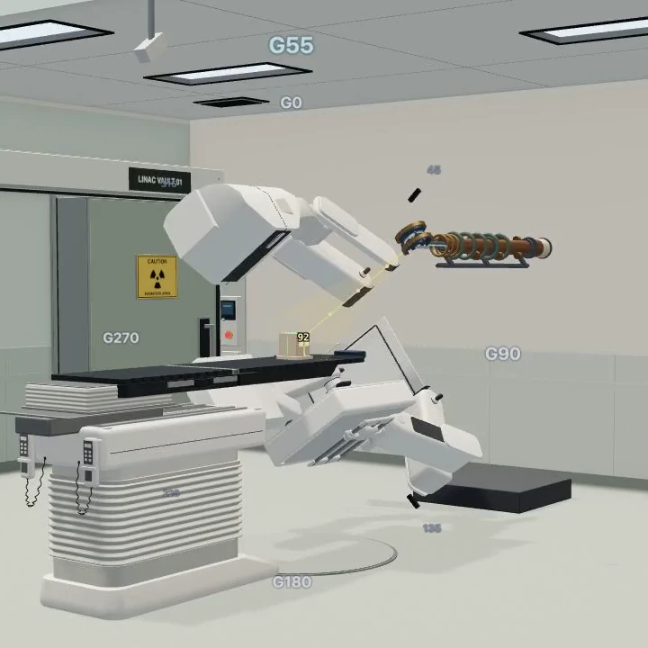
The October 13 tour starts with the complete native training linac and its whole internal assembly, then follows a continuous camera path to the electron gun, accelerating cavities, bending magnet and MLC banks before approaching a sectioned leaf. The camera settles before each demonstration. These are actual trainer WebGL captures; internal geometry is representative, and particle markers are enlarged and slowed for teaching.
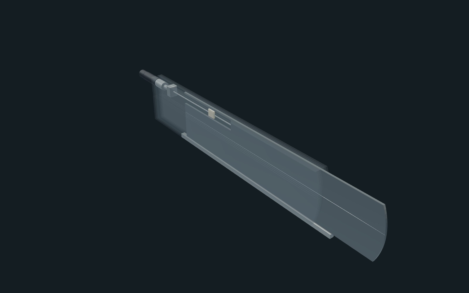
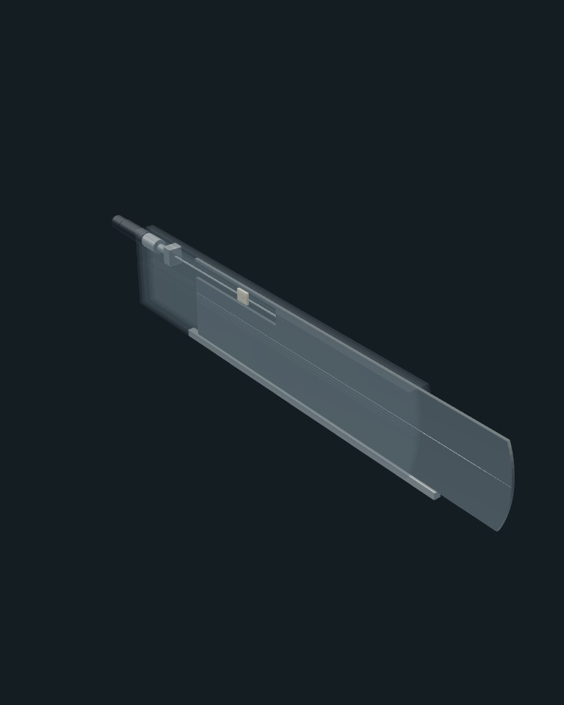
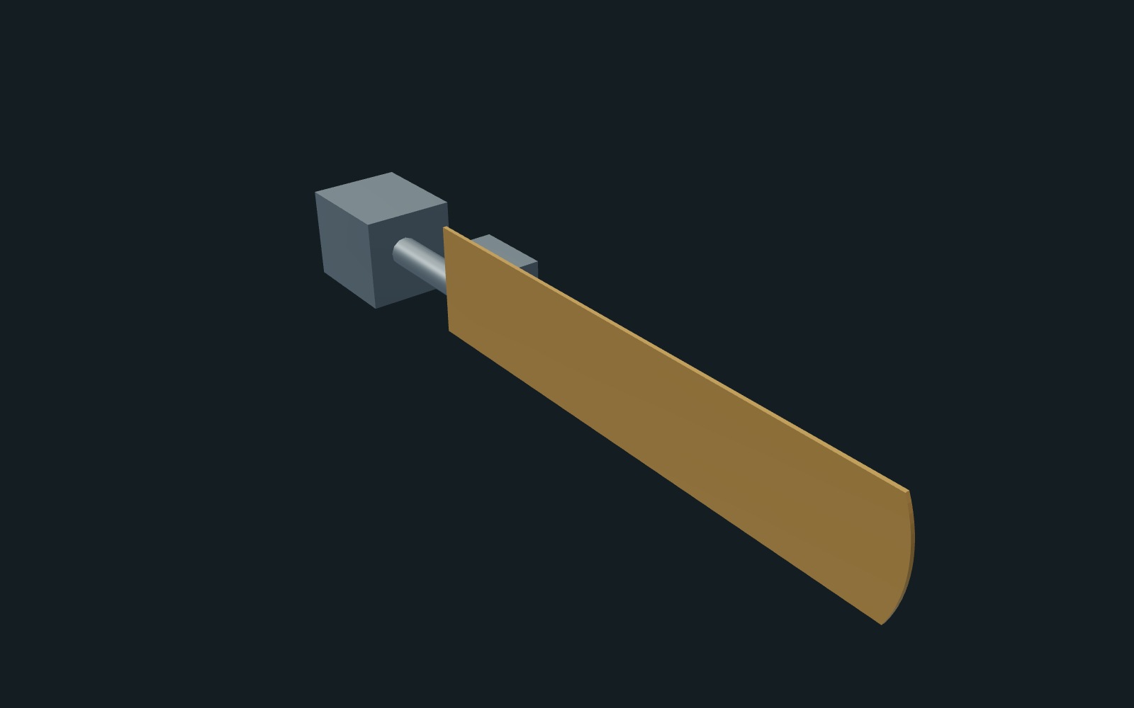
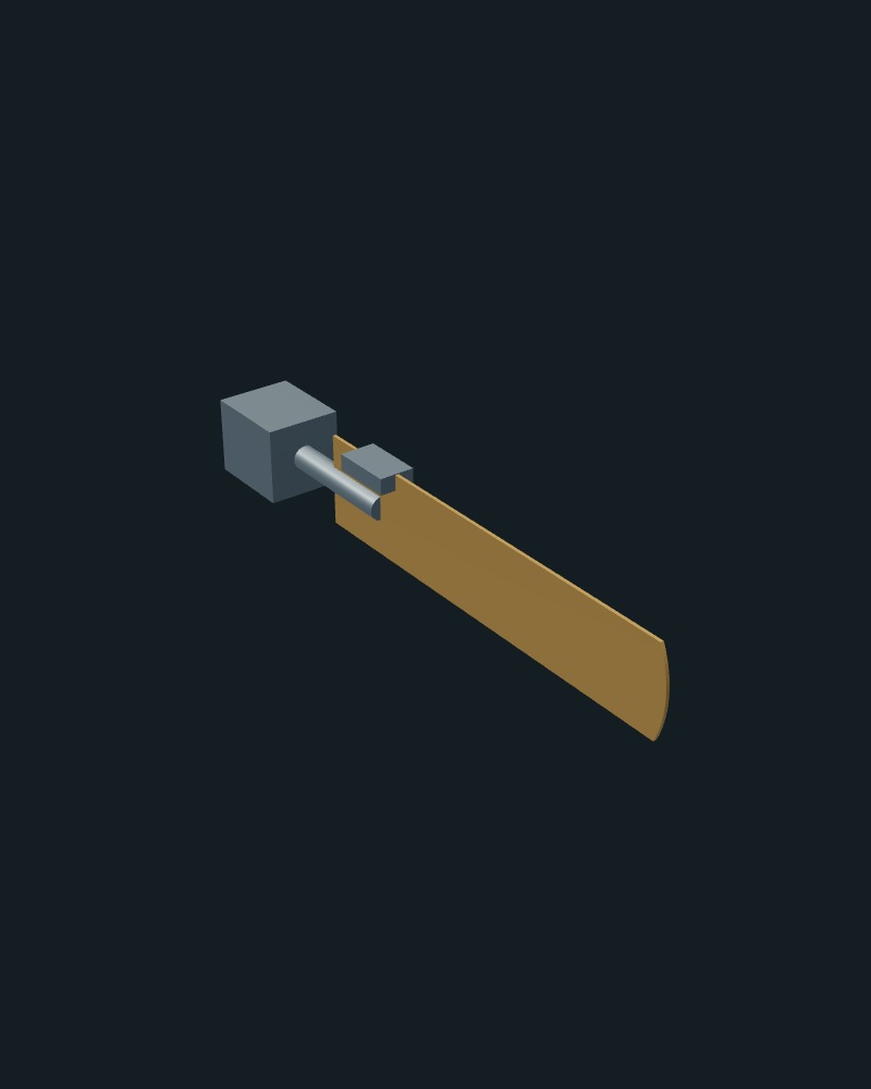
Camera, lighting, selection highlighting and temporary scene-copy opacity are staged locally. A gentle pullback and eased zoom connect neighboring native cutaways; the whole-machine context appears during the first approach and final return. Temporary material fades hand off between the parts. No image crossfades are used. The 27-second landscape and portrait films use 48 native frames per second for paused scroll seeking; the optional Play this part control loops only the stationary demonstration. Phone views use separate component framing. The shared MLC visual geometry was revised in Blender; the original 120-leaf arrangement, leaf-tip profile, treatment aperture and collision behavior are retained. The MLC chapter allows four seconds to establish the banks and approach the leaf before its two-second demonstration.
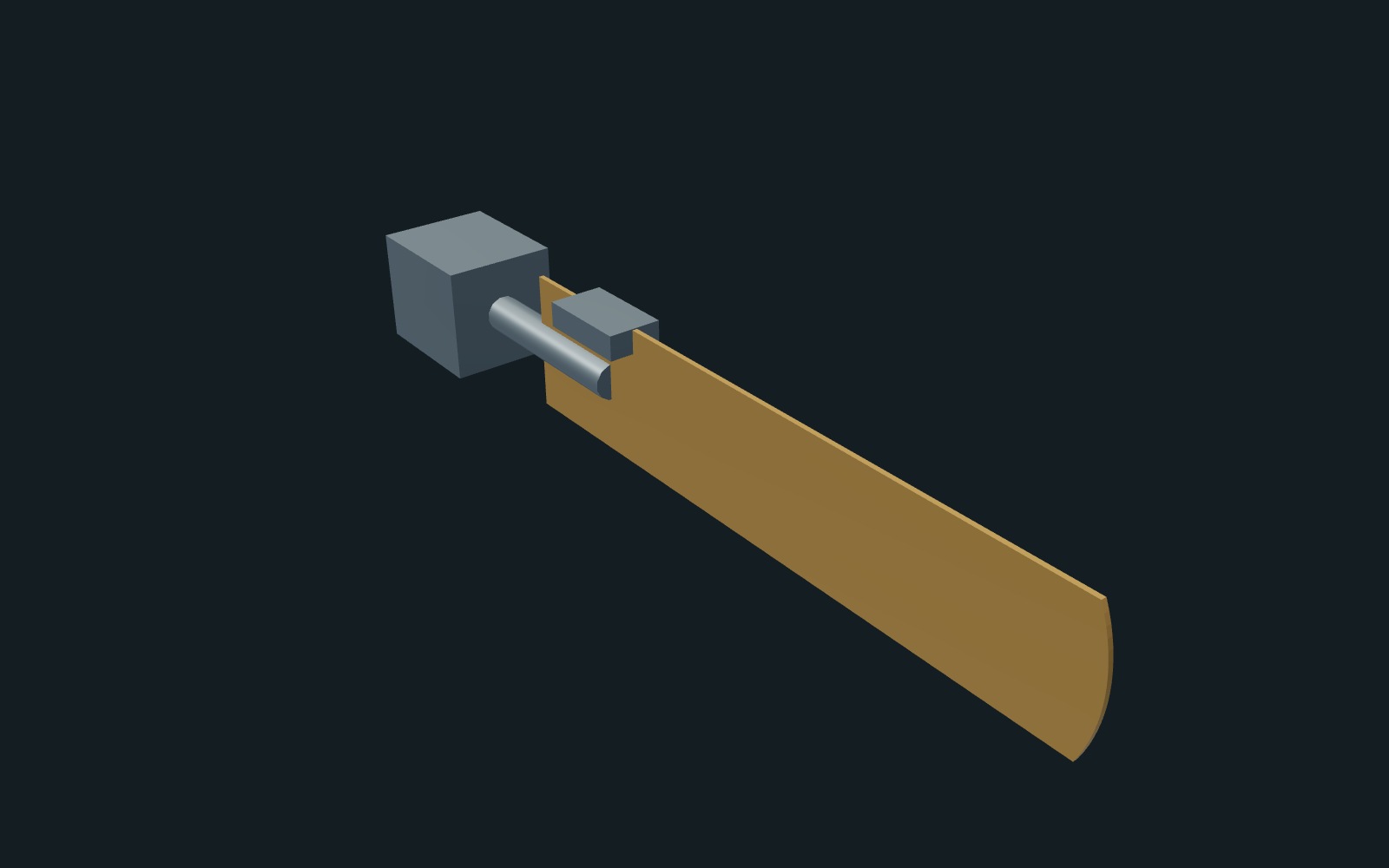
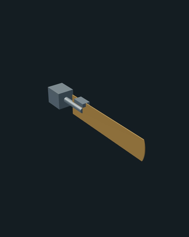
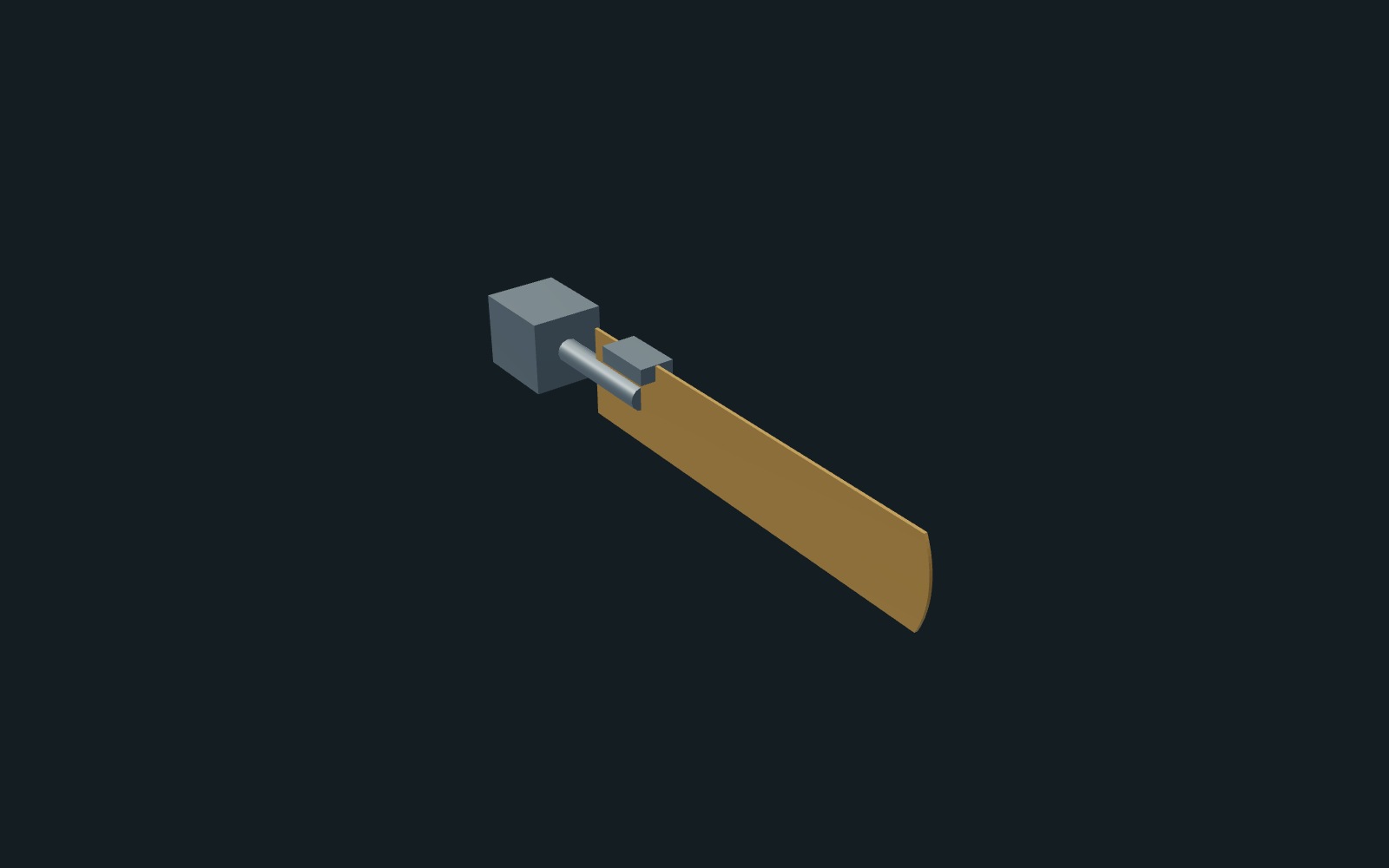
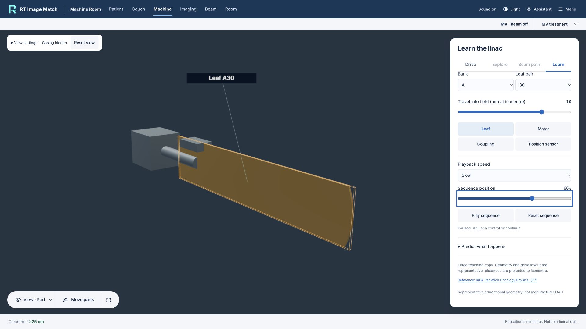
The MLC uses original Blender-authored leaves, guides, motor, coupler, threaded screw and translating nut, informed by AAPM Task Group 50 and Barnes et al. (2022). The service window, side relief, drive packaging, screw pitch and materials are illustrative, not manufacturer CAD or a dose calculation. The visible teaching copy does not move the treatment aperture.
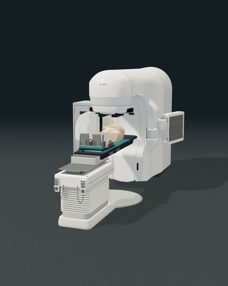
Whole-machine views retain the pelvic exterior from Prostate-Anatomical-Edge-Cases (TCIA), CC BY 4.0. No source head, facial field or private learner record is shown. Capture evidence and source hashes are recorded in docs/landing-linac-transitions-captures.json; exact media hashes are registered in docs/data-rights-register.json.
- linac-tour-outside-desktop-20261013-mlc.jpg
- linac-tour-internals-desktop-20261013-mlc.jpg
- linac-tour-gun-desktop-20261013-mlc.jpg
- linac-tour-cavities-desktop-20261013-mlc.jpg
- linac-tour-bend-desktop-20261013-mlc.jpg
- linac-tour-mlc-desktop-20261013-mlc.jpg
- linac-tour-return-desktop-20261013-mlc.jpg
- linac-tour-desktop-20261013-mlc.mp4
- linac-tour-outside-mobile-20261013-mlc.jpg
- linac-tour-internals-mobile-20261013-mlc.jpg
- linac-tour-gun-mobile-20261013-mlc.jpg
- linac-tour-cavities-mobile-20261013-mlc.jpg
- linac-tour-bend-mobile-20261013-mlc.jpg
- linac-tour-mlc-mobile-20261013-mlc.jpg
- linac-tour-return-mobile-20261013-mlc.jpg
- linac-tour-mobile-20261013-mlc.mp4
October 12 component-transition captures
The earlier 25-second film used the previous single-leaf visual. Its exact source and media hashes remain in docs/landing-linac-transitions-20261012-captures.json.
- linac-tour-outside-desktop-20261012-v3.jpg
- linac-tour-internals-desktop-20261012-v3.jpg
- linac-tour-gun-desktop-20261012-v3.jpg
- linac-tour-cavities-desktop-20261012-v3.jpg
- linac-tour-bend-desktop-20261012-v3.jpg
- linac-tour-mlc-desktop-20261012-v3.jpg
- linac-tour-return-desktop-20261012-v3.jpg
- linac-tour-desktop-20261012-v3.mp4
- linac-tour-outside-mobile-20261012-v3.jpg
- linac-tour-internals-mobile-20261012-v3.jpg
- linac-tour-gun-mobile-20261012-v3.jpg
- linac-tour-cavities-mobile-20261012-v3.jpg
- linac-tour-bend-mobile-20261012-v3.jpg
- linac-tour-mlc-mobile-20261012-v3.jpg
- linac-tour-return-mobile-20261012-v3.jpg
- linac-tour-mobile-20261012-v3.mp4
Earlier October 12 connected-camera captures
The previous 24 fps tour retained whole-machine context between component close-ups. Its source and camera evidence remains in docs/landing-linac-refined-captures.json.
- linac-tour-outside-desktop-20261012-v2.jpg
- linac-tour-internals-desktop-20261012-v2.jpg
- linac-tour-gun-desktop-20261012-v2.jpg
- linac-tour-cavities-desktop-20261012-v2.jpg
- linac-tour-bend-desktop-20261012-v2.jpg
- linac-tour-mlc-desktop-20261012-v2.jpg
- linac-tour-return-desktop-20261012-v2.jpg
- linac-tour-desktop-20261012-v2.mp4
- linac-tour-outside-mobile-20261012-v2.jpg
- linac-tour-internals-mobile-20261012-v2.jpg
- linac-tour-gun-mobile-20261012-v2.jpg
- linac-tour-cavities-mobile-20261012-v2.jpg
- linac-tour-bend-mobile-20261012-v2.jpg
- linac-tour-mlc-mobile-20261012-v2.jpg
- linac-tour-return-mobile-20261012-v2.jpg
- linac-tour-mobile-20261012-v2.mp4
Earlier October 12 captures
The earlier tour used native-frame dissolves. Its source and camera evidence remains in docs/landing-linac-internals-captures.json.
- linac-tour-outside-desktop-20261012.jpg
- linac-tour-internals-desktop-20261012.jpg
- linac-tour-gun-desktop-20261012.jpg
- linac-tour-cavities-desktop-20261012.jpg
- linac-tour-bend-desktop-20261012.jpg
- linac-tour-mlc-desktop-20261012.jpg
- linac-tour-return-desktop-20261012.jpg
- linac-tour-desktop-20261012.mp4
- linac-tour-outside-mobile-20261012.jpg
- linac-tour-internals-mobile-20261012.jpg
- linac-tour-gun-mobile-20261012.jpg
- linac-tour-cavities-mobile-20261012.jpg
- linac-tour-bend-mobile-20261012.jpg
- linac-tour-mlc-mobile-20261012.jpg
- linac-tour-return-mobile-20261012.jpg
- linac-tour-mobile-20261012.mp4
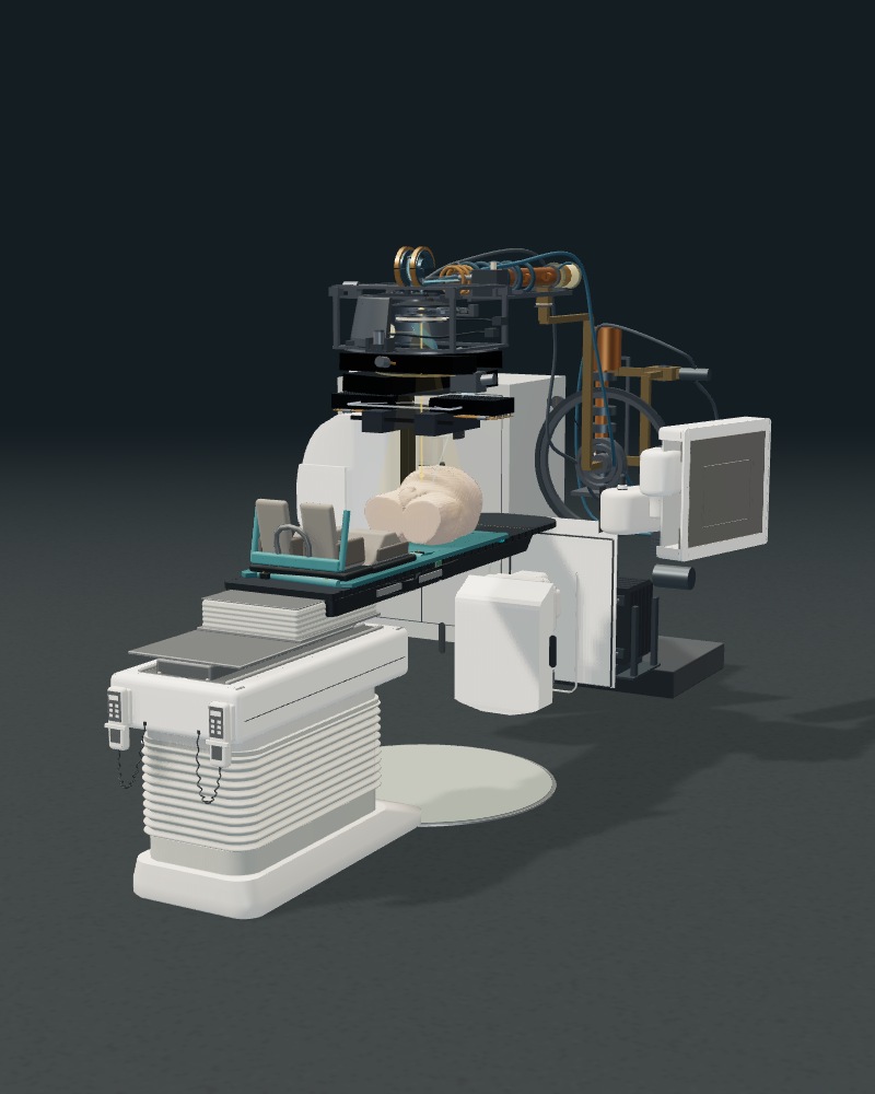
Machine Room: October 11 scroll tour
The landing tour uses new native recordings of the trainer’s representative linac, electron gun, accelerating cavities, bending magnet and single-leaf MLC study. The original teaching models supply the enlarged, slowed electron/RF markers and separate moving leaf copy. These are educational illustrations, not manufacturer CAD, calibrated particle transport, machine timing or dose calculations. The leaf study does not move the actual treatment aperture.
The camera moves, lighting, background and short native-frame dissolves are staged only in the local capture fixture. No machine geometry or protected model bytes are changed. Separate landscape and portrait films are 22 seconds at 24 frames per second; scroll position controls their paused playheads. The complete-machine views include the existing pelvic exterior from Prostate-Anatomical-Edge-Cases (TCIA), licensed CC BY 4.0; the full citation appears below. Close-ups show first-party teaching geometry. No source head, facial field or private learner record is shown. Source/model hashes, camera settings and exact media hashes are in docs/landing-linac-tour-captures.json and docs/data-rights-register.json.
- linac-tour-outside-desktop-20261011.jpg
- linac-tour-gun-desktop-20261011.jpg
- linac-tour-cavities-desktop-20261011.jpg
- linac-tour-bend-desktop-20261011.jpg
- linac-tour-mlc-desktop-20261011.jpg
- linac-tour-return-desktop-20261011.jpg
- linac-tour-desktop-20261011.mp4
- linac-tour-outside-mobile-20261011.jpg
- linac-tour-gun-mobile-20261011.jpg
- linac-tour-cavities-mobile-20261011.jpg
- linac-tour-bend-mobile-20261011.jpg
- linac-tour-mlc-mobile-20261011.jpg
- linac-tour-return-mobile-20261011.jpg
- linac-tour-mobile-20261011.mp4
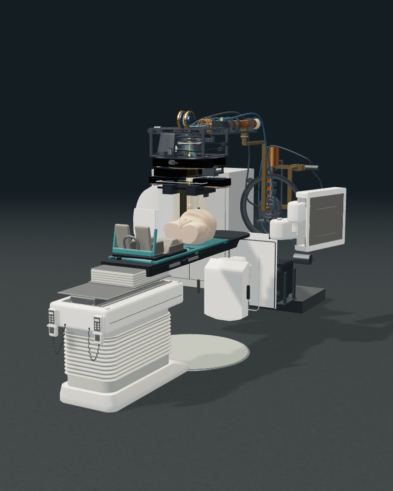
Machine Room: archived October 10 presentation captures
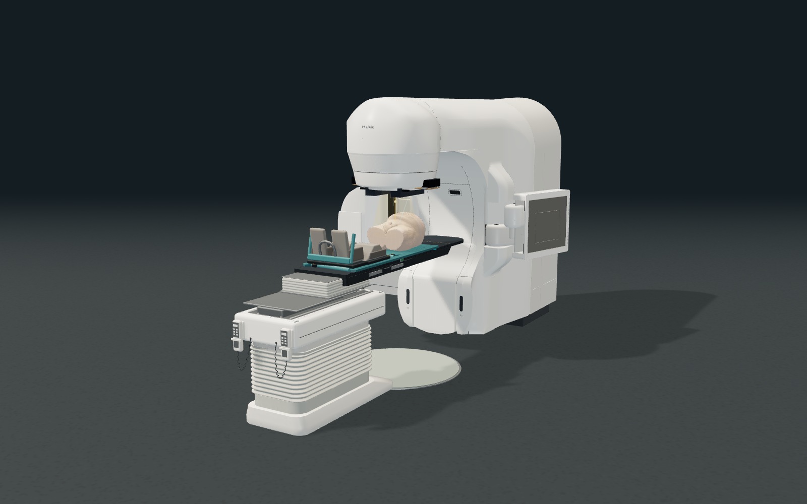
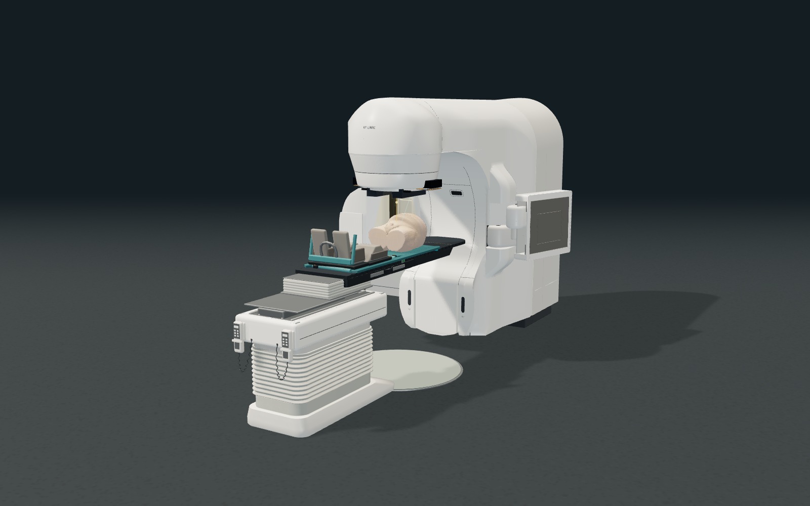
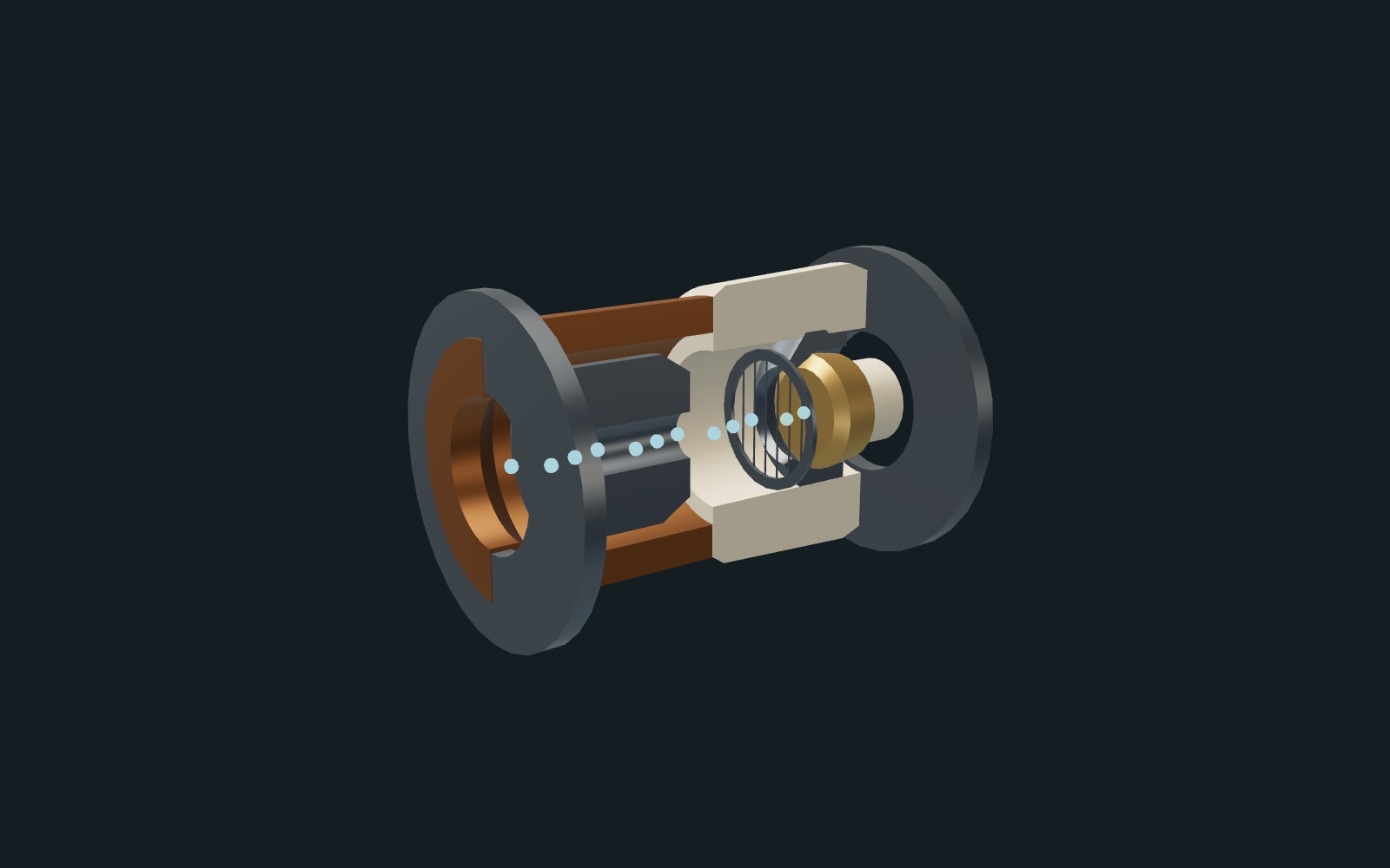
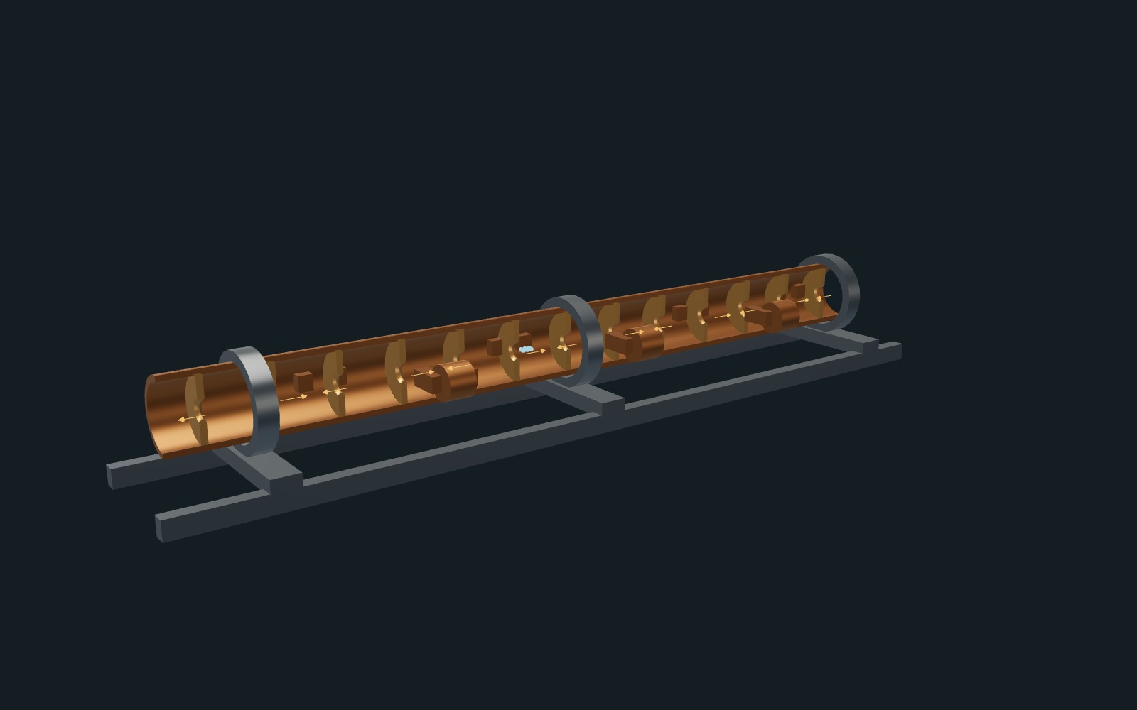
The landing-page linac uses fresh native frames from the current trainer model, captured on October 10, 2026. Exterior and internal views share the same camera path, with separate landscape and portrait compositions. The interface, labels, optical lamps and room decoration are hidden for these presentation captures. Background, floor color and existing lights are adjusted; the floor is extended and distance haze softens its horizon. Machine, couch, support and pelvic exterior geometry are unchanged. These are staged simulator views, not photographs of clinical equipment.
The visible pelvic exterior uses Thompson, Reid F., et al. (2023), Prostate-Anatomical-Edge-Cases, Version 1 (TCIA), CC BY 4.0. Its finite source extent is retained. No source head or facial field is shown. Equipment and internal components are representative teaching geometry, independent of manufacturers. Capture takes place in the local fictional-account fixture without private learner records.
The silent eight-second loops encode native camera samples at 24 frames per second, without generated imagery or frame interpolation. They are presentation recordings, not real-time workflow demonstrations. Motion pauses outside the viewport and a still image remains available for reduced motion, data-saving preferences or playback failure. Source revision, model hash, camera settings and exact media hashes are recorded in docs/landing-linac-captures.json and docs/data-rights-register.json.
- linac-outside-desktop-20261010.jpg
- linac-outside-desktop-20261010.mp4
- linac-inside-desktop-20261010.jpg
- linac-inside-desktop-20261010.mp4
- linac-outside-mobile-20261010.jpg
- linac-outside-mobile-20261010.mp4
- linac-inside-mobile-20261010.jpg
- linac-inside-mobile-20261010.mp4
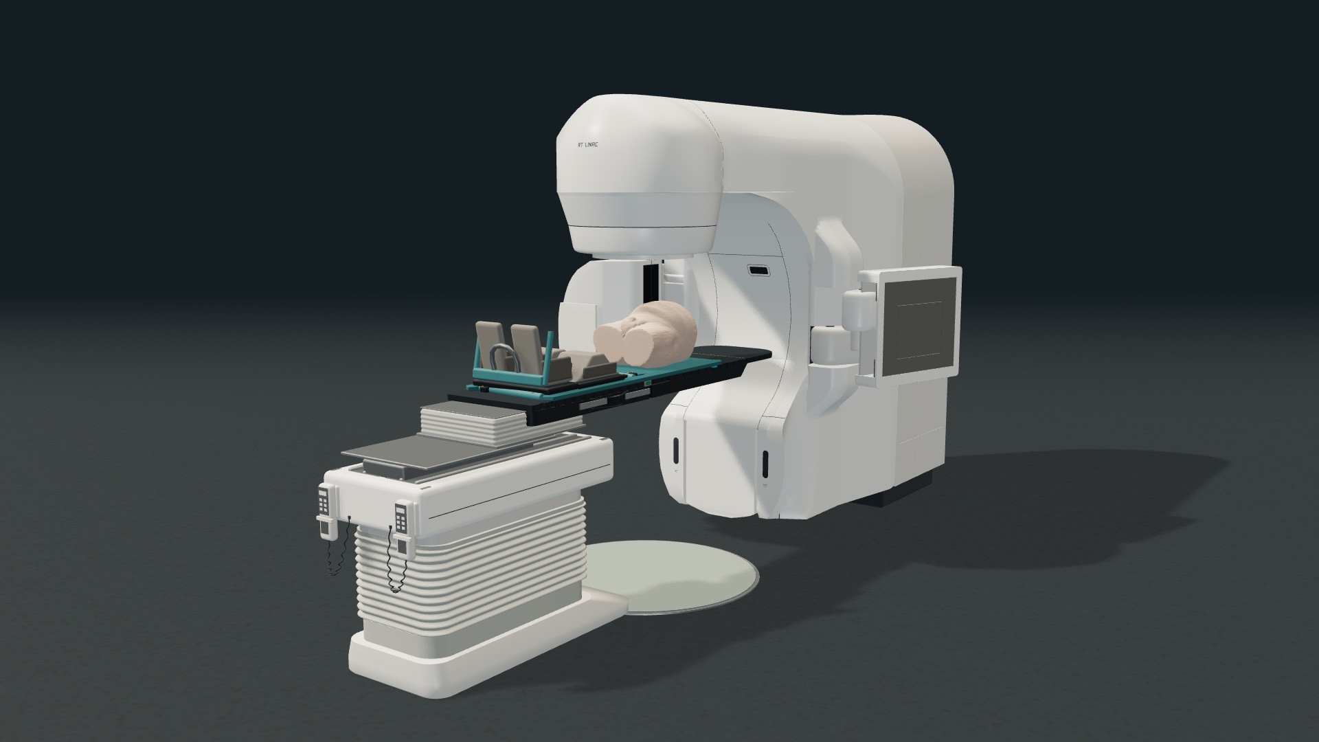
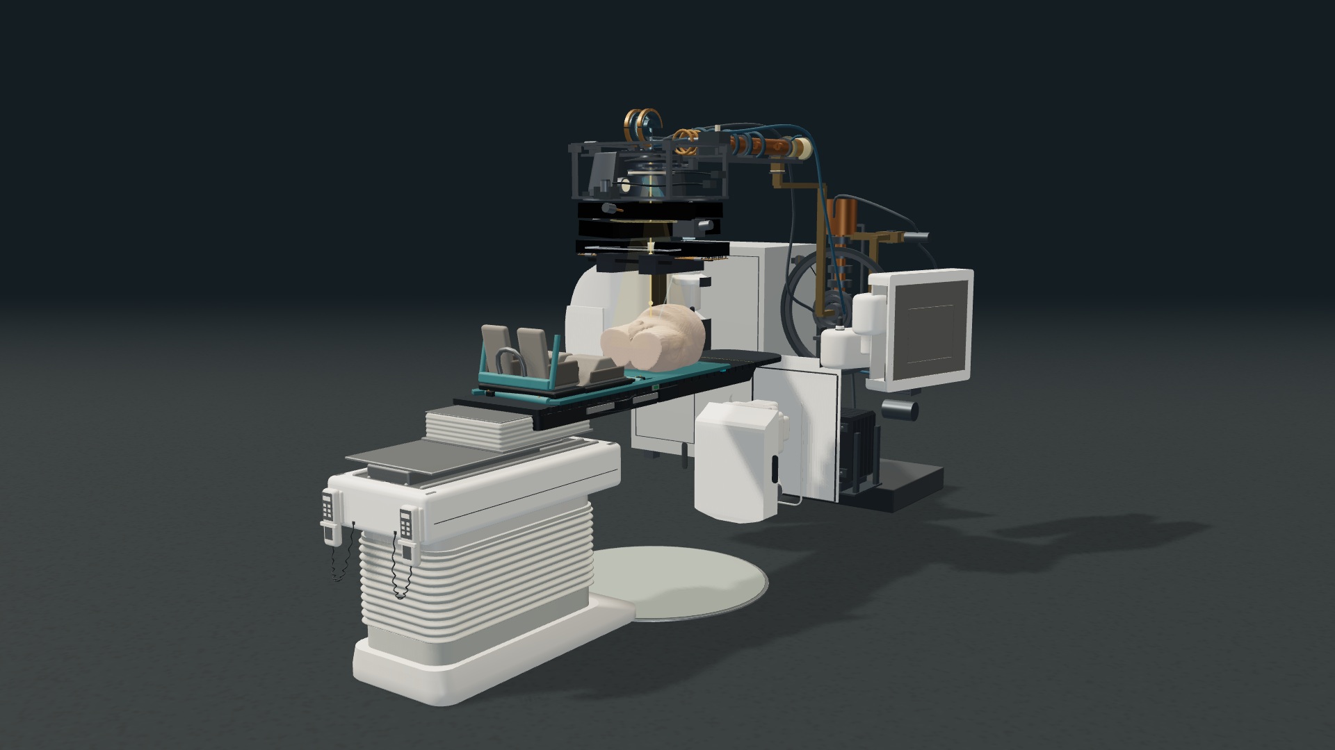
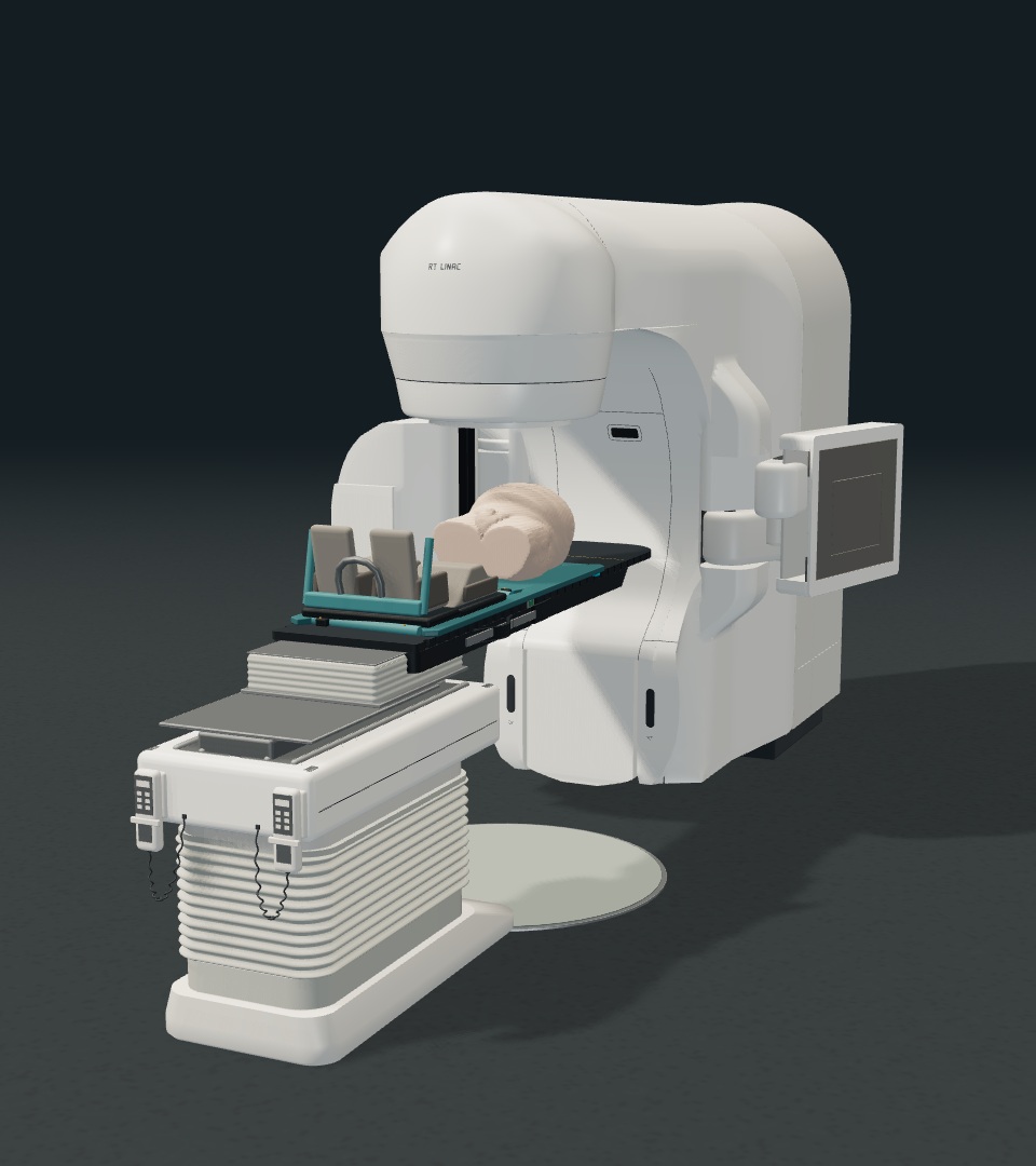
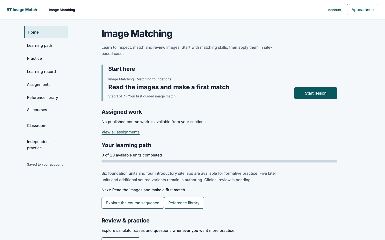
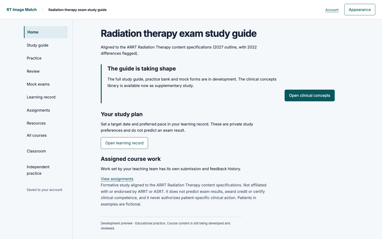
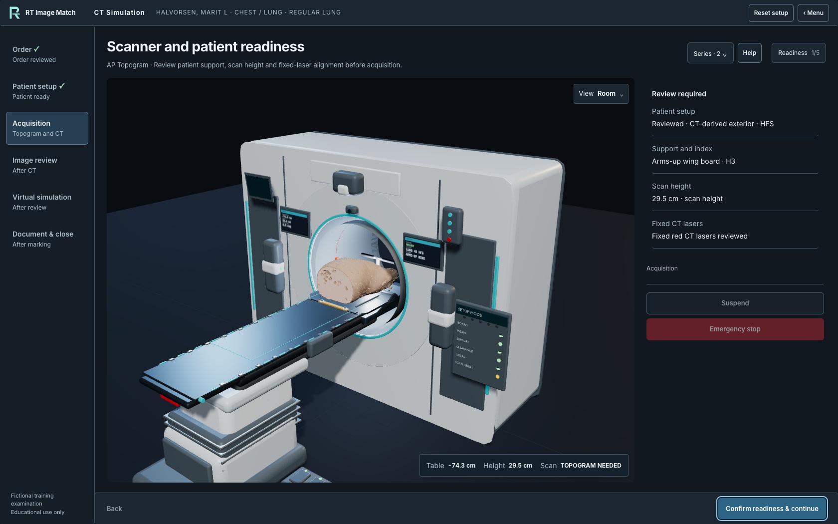
Cinematic preview: courses and CT Simulation
Five additional native screenshots were captured on October 9, 2026. Image Matching and Radiation Therapy Study Guide images show the actual development applications with the existing local fictional-account fixture and empty learner records. They contain no patient image or real learner record. Both courses remain in development.
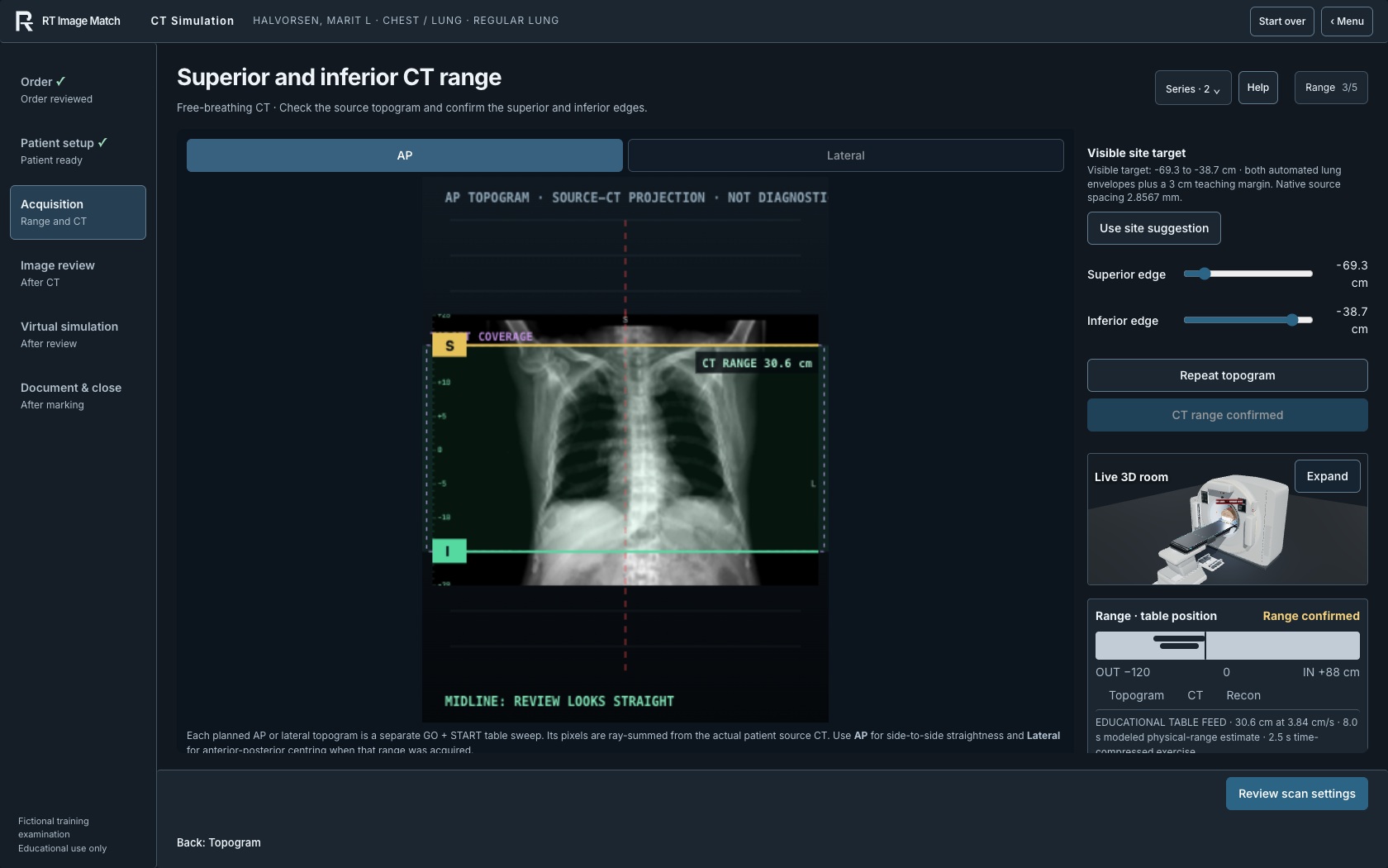
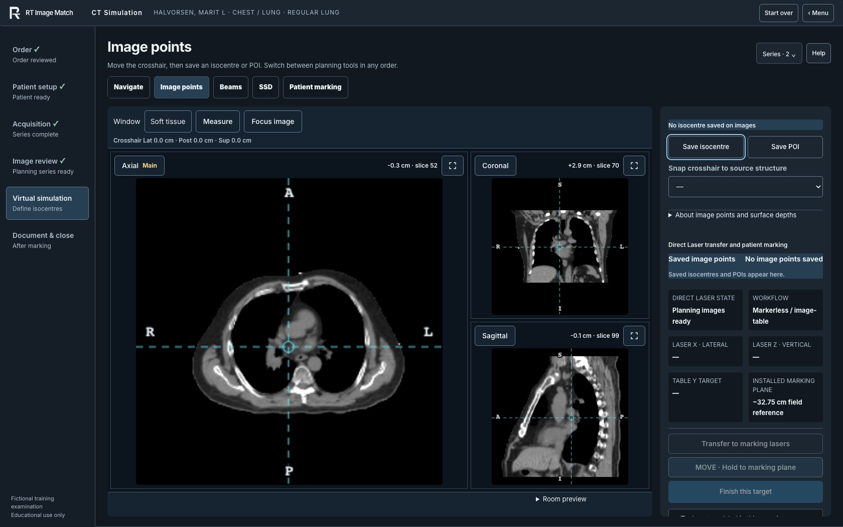
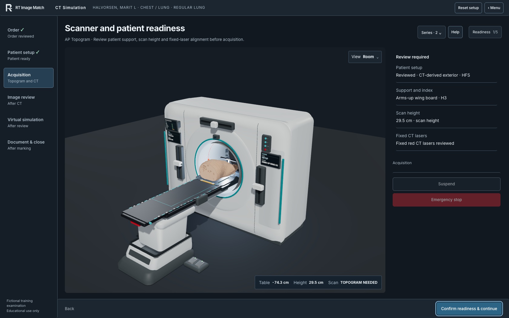
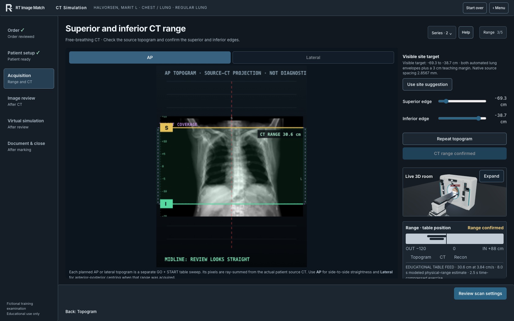
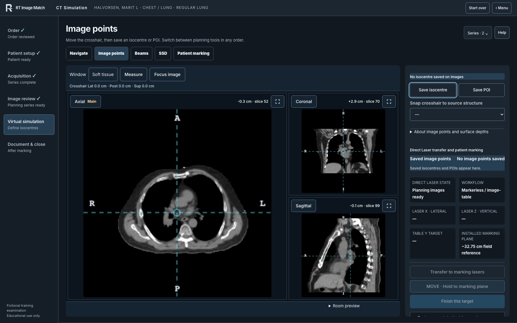
The October 9 CT Simulation images show the captured preview interface for the Regular Lung workflow. Source CT projections, multiplanar images and the finite arms-up BODY-derived exterior use the 4D-Lung collection, CC BY 3.0, with the full citation below. The exterior uses a matte synthetic color and no source texture; acquired caps are retained without anatomical continuation. No complete head or facial field is shown. Scanner and support geometry are first-party educational models, independent of manufacturers.
The CT capture uses the established local owner fixture. Its existing completion helpers advance topogram and CT timing to display later review and virtual-simulation states; these are fictional training records, not performed clinical examinations. The displayed patient name is fictional. Source pixels and product UI are unchanged. Only still images are public: source volumes, the CT model and owner-only access remain protected. File hashes, source revisions and capture timestamps are recorded in docs/landing-cinematic-expansion-captures.json.
- cinematic-course-matching-20261009.jpg
- cinematic-course-registry-20261009.jpg
- cinematic-ct-room-20261009.jpg
- cinematic-ct-imaging-20261009.jpg
- cinematic-ct-virtual-20261009.jpg
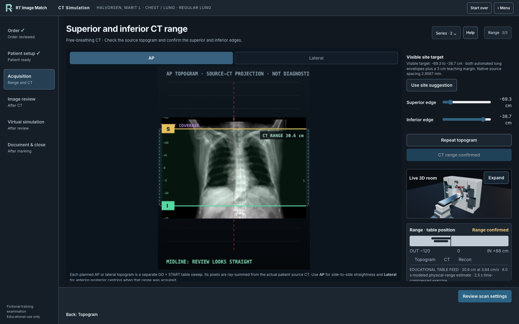
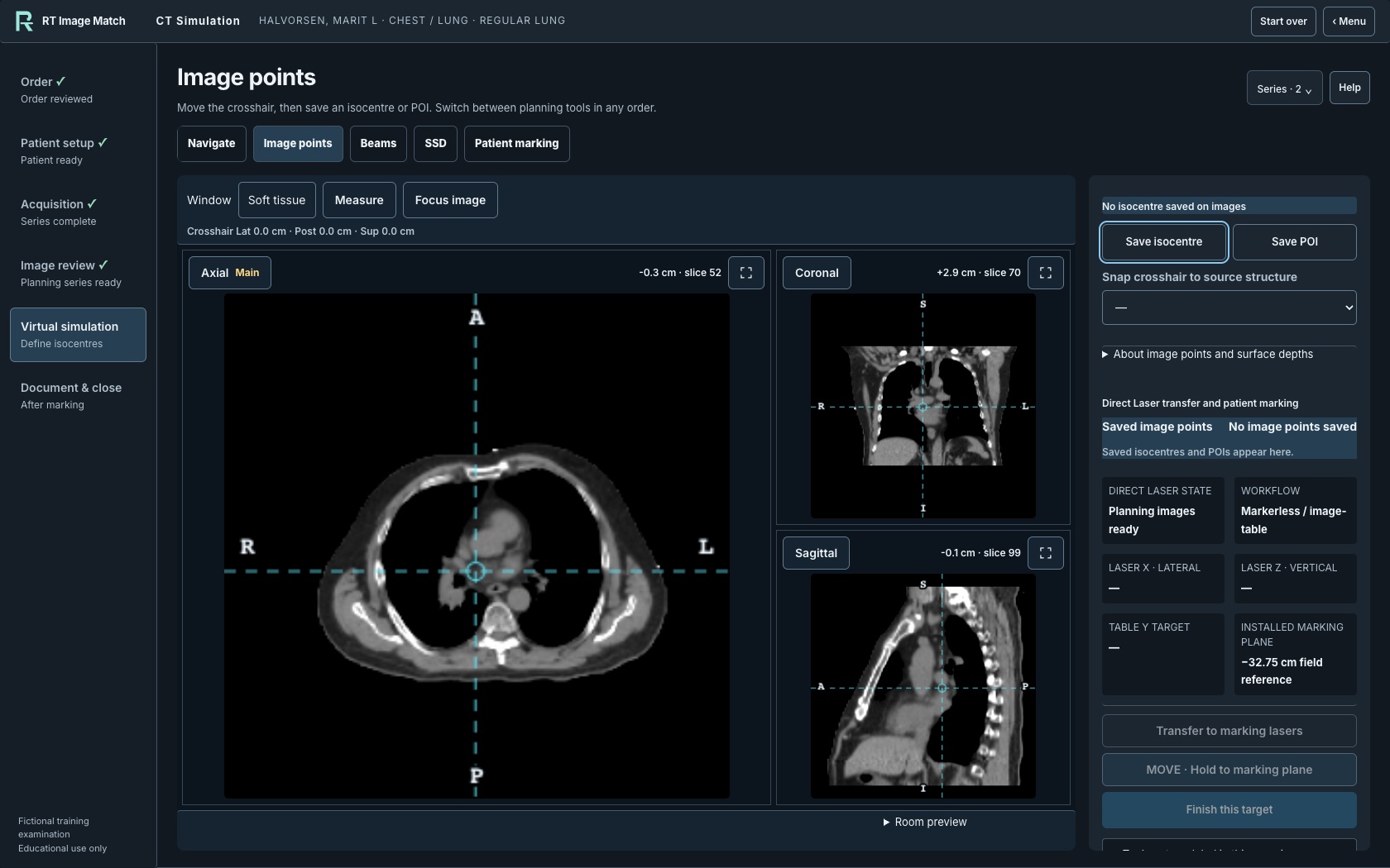
CT scanner: October 10 presentation captures
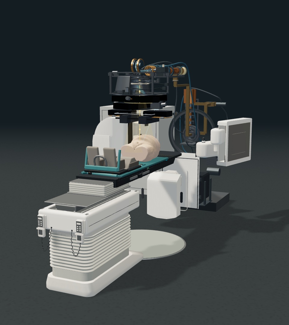
The landing page's first CT image and the trainer CT card show the current simulator model with an empty table, captured in the local fictional-account fixture on October 10, 2026. Separate landscape and portrait cameras frame the complete scanner, table and floor base. The interface and teaching guides are hidden. Background, floor tint, camera and existing lights are adjusted for presentation; 64 native lighting samples soften the shadows. These are staged simulator views, not photographs of clinical equipment.
The bare table uses the existing renderer's positioning controls. Machine geometry and materials are unchanged, and no patient anatomy is selected or displayed. No generated imagery, pixel retouching, manufacturer CAD or private learner record is used. The equipment is first-party representative teaching geometry, independent of manufacturers. Source and generator hashes, the staged pose, lighting, camera settings and exact image hashes are recorded in docs/landing-ct-presentation-captures.json and docs/data-rights-register.json. The topogram and virtual-simulation screenshots retain the 4D-Lung attribution below.
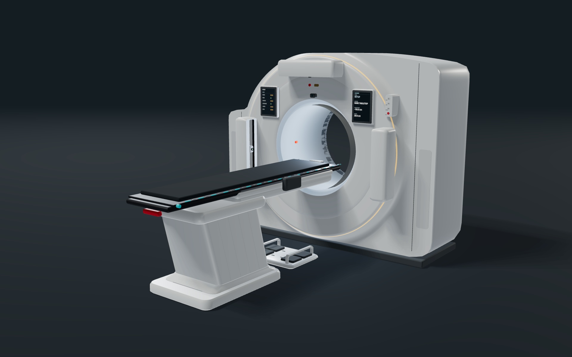
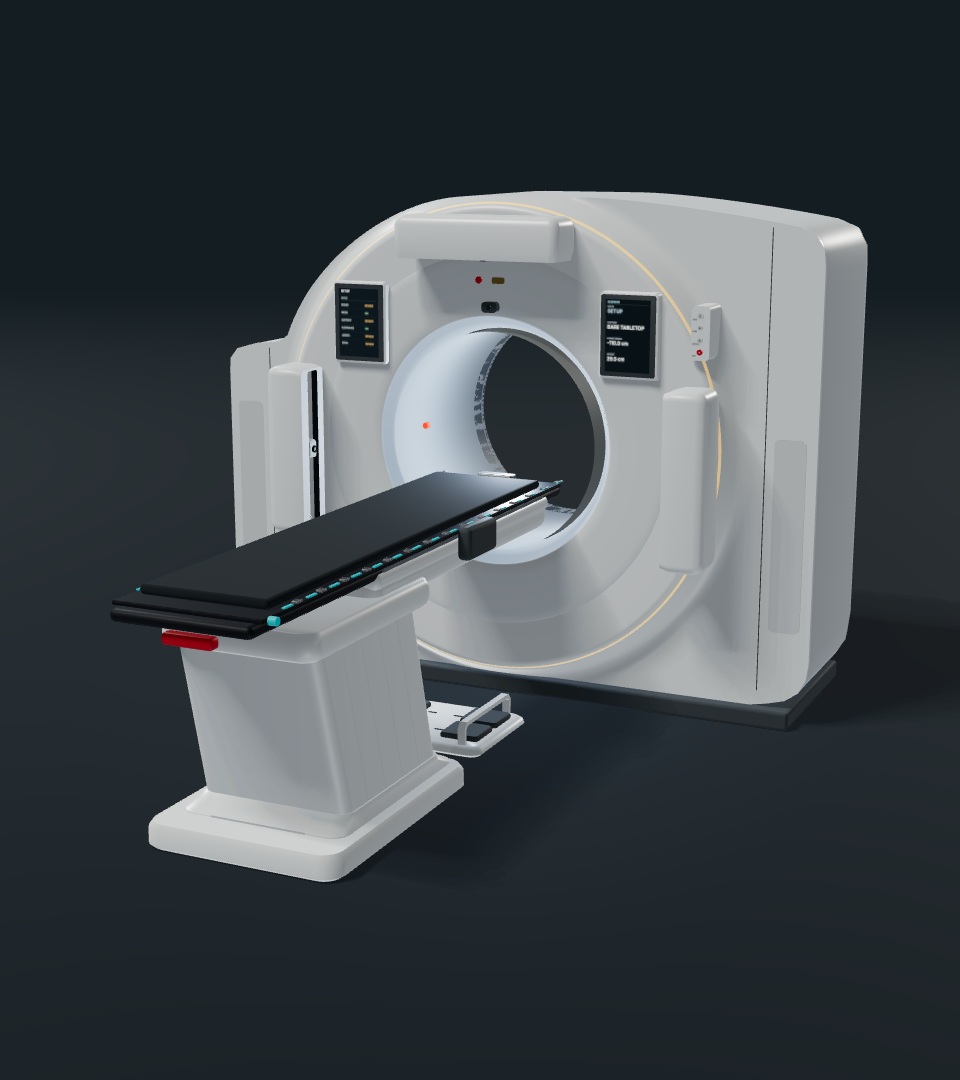
Updated CT scanner and table-base captures
The CT workflow screenshots were captured on October 10, 2026, with the updated circular scanner casing, enclosed laser guides, portrait tablets, slim remote, camera and halo indicators, outer moodlight, guarded footswitch, inclined table enclosure, white carriage, fine side joints and low floor base. The protective-cover motion is an independently authored visual proxy; hidden mechanics and enclosure proportions are not manufacturer specifications. The retained room screenshot uses the ordinary camera zoom control to show the complete base. The landing page's topogram and virtual-simulation views were refreshed in the same local fictional-account session. These are simulator screenshots, not photographs of clinical equipment.
The images retain the Regular Lung workflow and the 4D-Lung collection attribution and CC BY 3.0 license described above. No source head or facial field is shown. Existing fixture completion helpers prepare the later imaging states; displayed names and examination values are fictional. No generated anatomy or interface is added. Exact model, renderer and controller hashes, source revision and capture timestamps are recorded in docs/landing-ct-scanner-captures.json. The October 9 captures remain available in the preceding archive. The two earlier October 10 sets are retained below, with their source hashes in docs/landing-ct-scanner-captures-20261010-r1.json and docs/landing-ct-scanner-captures-20261010-r2.json.
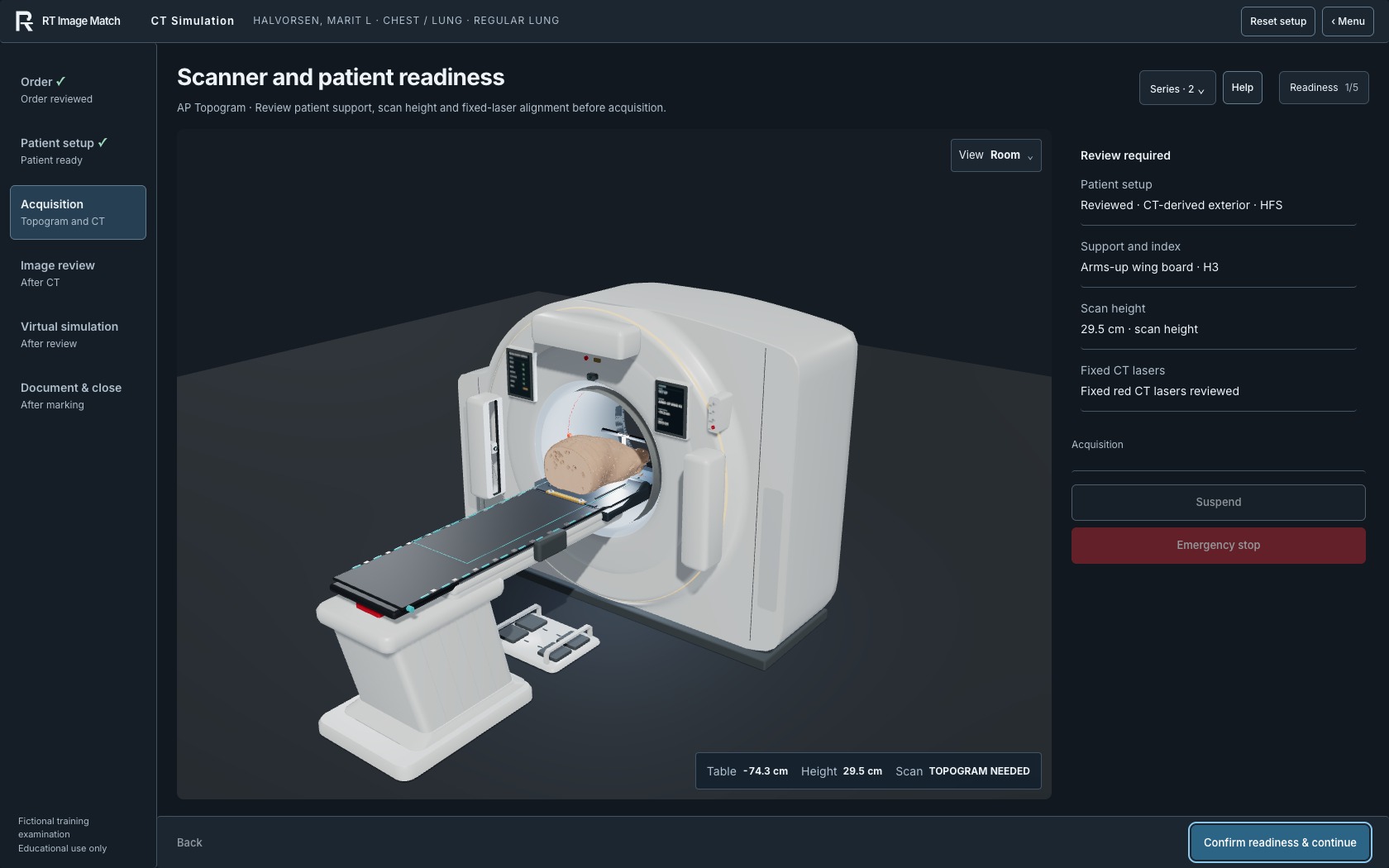
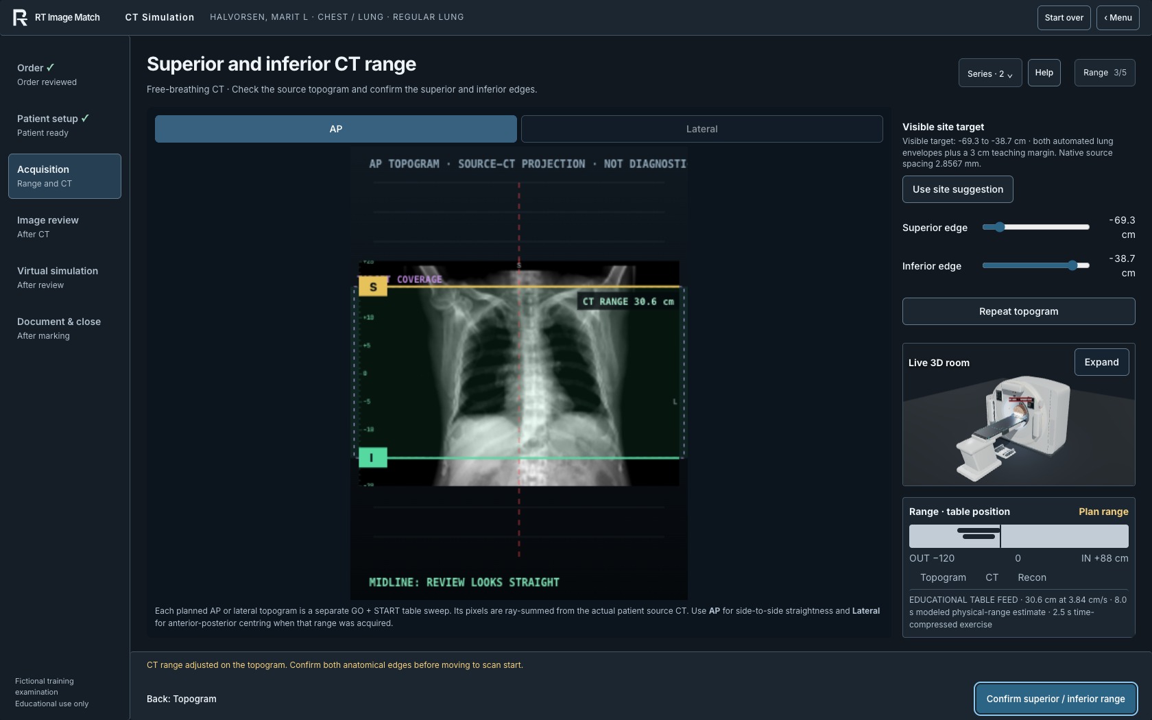
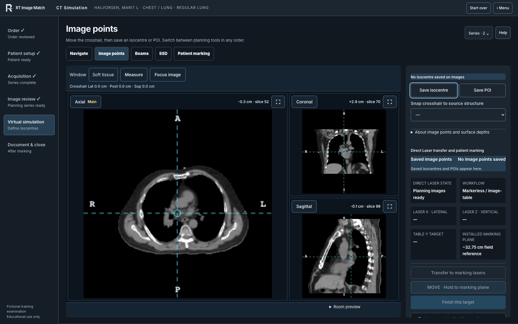
Archived second October 10 set: scanner room, topogram, virtual simulation.
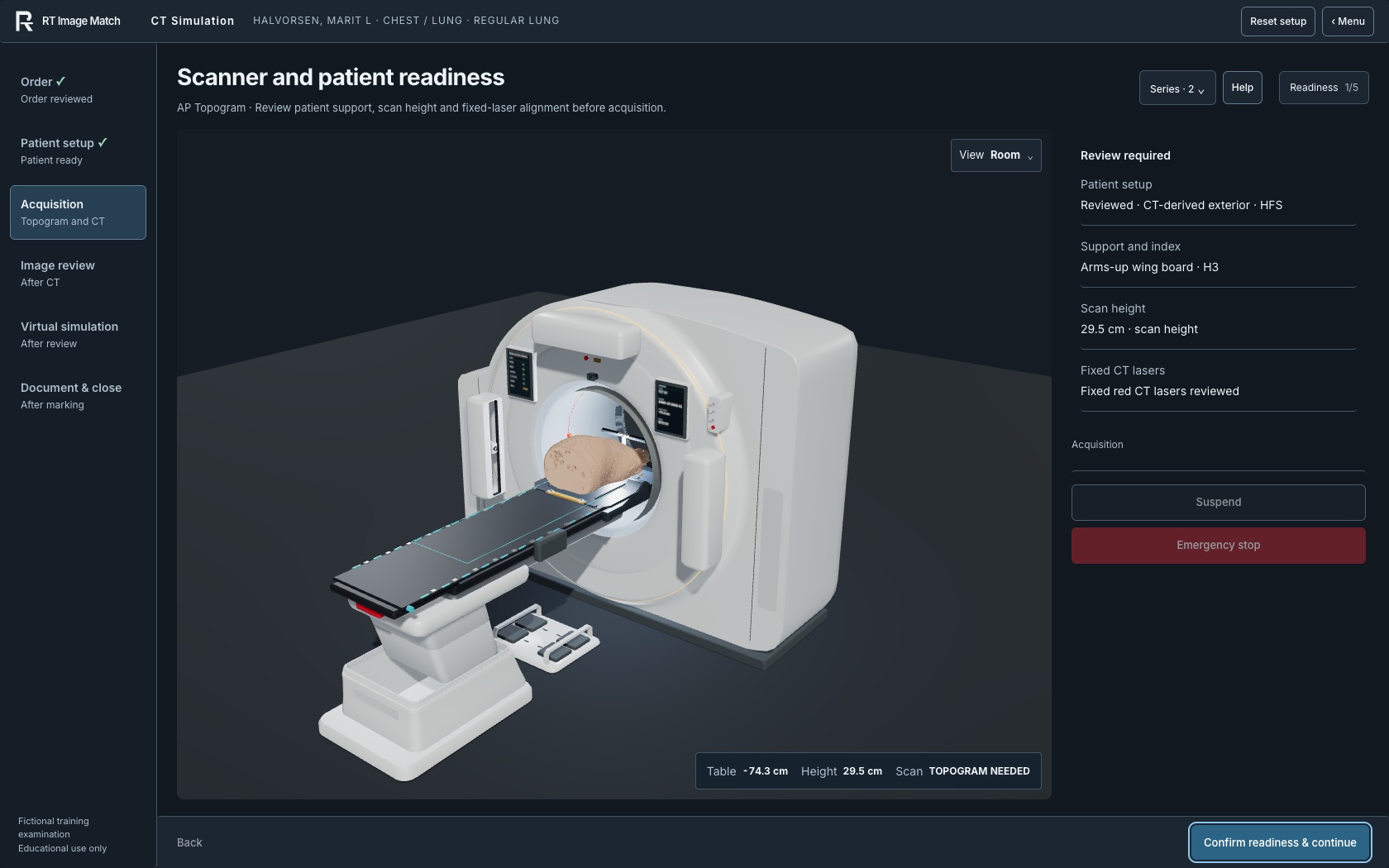
Archived first October 10 set: scanner room, topogram, virtual simulation.
Landing-page videos and captures
RT Image Match — Radiation therapy training
The current 2 minute 24 second film shows image registration, a prostate treatment workflow, DIBH coaching preparation, LINAC anatomy, the case library and Classroom teaching tools. September 30 fictional-account recordings are combined with October 5 live-trainer and fictional Classroom captures. This 24-scene revision adds three fresh recordings of registration, MLC mechanics and natural typing in the Weekly planner. Closer framing, one fictional treatment case with stage indicators and longer verification, DIBH and results passages retain the complete 2:24 feature coverage. Every scene preserves consecutive encoded frames at 25 fps, with brief reading holds and natural UI pauses; sequential browser capture is time-compressed and is not a claim of real-time 25 fps acquisition.
Native Higgsedit titles and framing sit outside the recorded software views. No source anatomy, displayed values or intermediate movement is generated, redrawn or interpolated. Independent matching is ungraded practice. Full Treatment shows the beginning of a VMAT arc, not a completed fraction. DIBH uses a local synthetic transcript fixture for coaching preparation; voice never starts imaging or delivery. Classroom names, schedules, attendance and results are fictional. The equipment uses representative teaching geometry, not manufacturer CAD, and has no clinical dose model. No source head or facial imaging, private learner records, account details or microphone recordings appear. One 1920 × 1080 landscape MP4 serves every screen. Educational use only; not for clinical decisions.
Prostate CBCT and Full Treatment: Thompson, Reid F., et al. (2023), Prostate-Anatomical-Edge-Cases, Version 1 (TCIA), CC BY 4.0. Planning-derived educational images use simulated daily transforms and overlays. Pelvis 2D/2D: Zuley, Margarita L., et al. (2016), TCGA-PRAD, Version 4 (TCIA), CC BY 3.0. Planning DRRs and simulated setup images are trimmed and proportionally scaled. The results shown here are in whole or part based upon data generated by the TCGA Research Network: cancer.gov/tcga.
Partial Breast L reference: Armato III, Samuel G., et al. (2015), LIDC-IDRI, Version 4 (TCIA), CC BY 3.0. The film frames the simulated room and respiratory monitor. Full collection citations and TCIA usage restrictions apply.
Music: “Inspired” by Kevin MacLeod (incompetech.com), licensed under Creative Commons Attribution 4.0. The excerpt runs from source time 32 to 176 seconds, with loudness normalization and entrance/ending fades; speed and pitch are unchanged. All capture audio is discarded. No speech, trainer sounds or microphone recordings are included. Typography uses Inter under the SIL Open Font License. No source author or organization is implied to endorse the film.
Fresh software screenshots
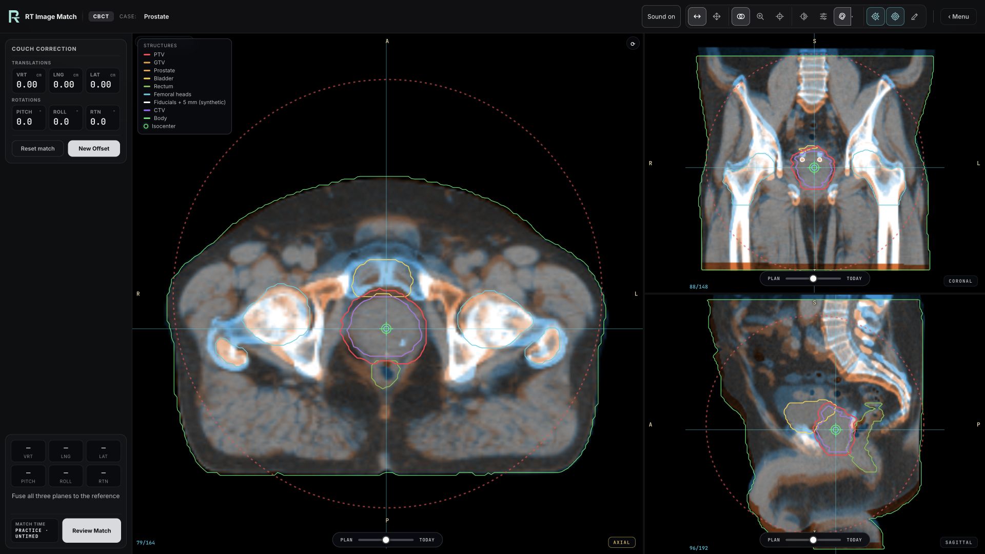
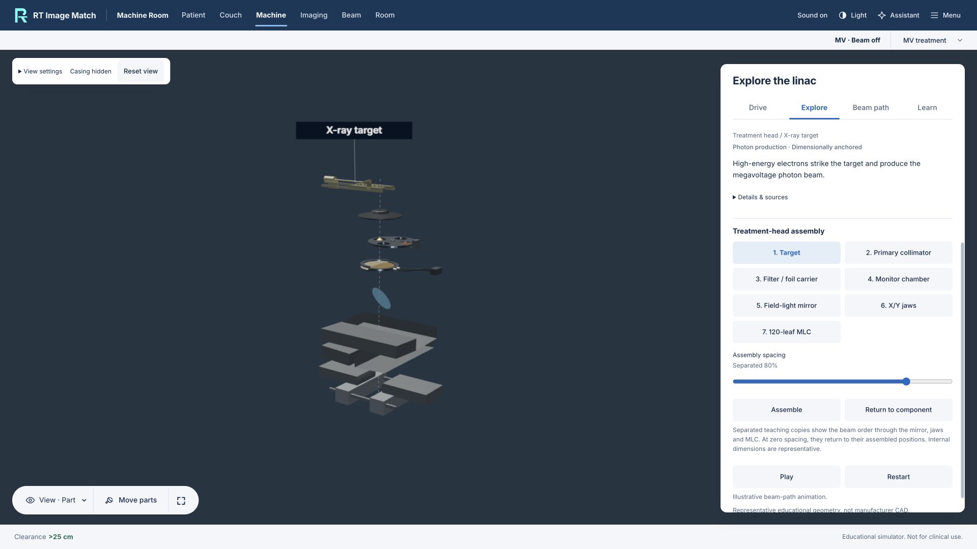
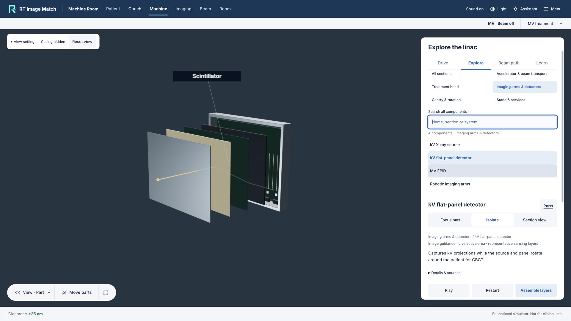
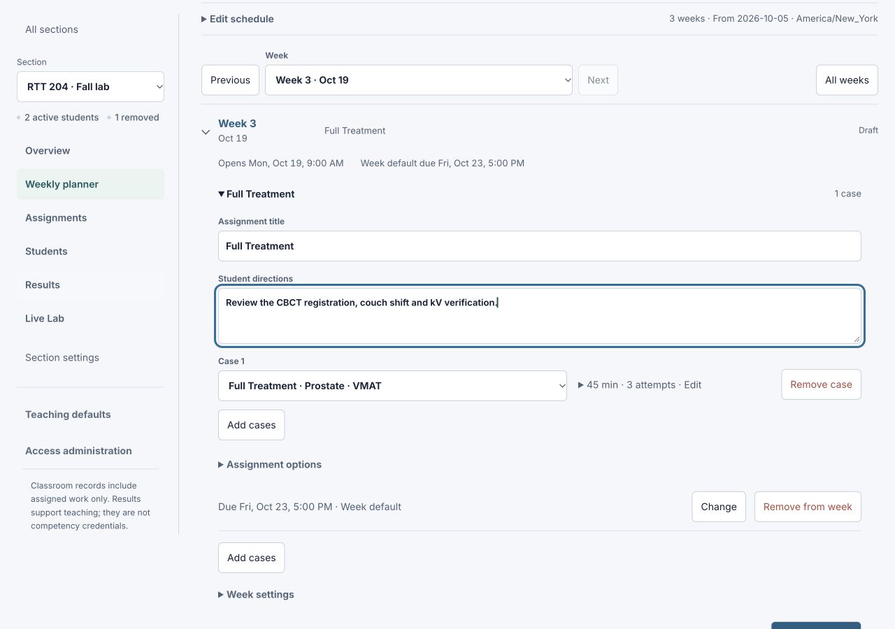
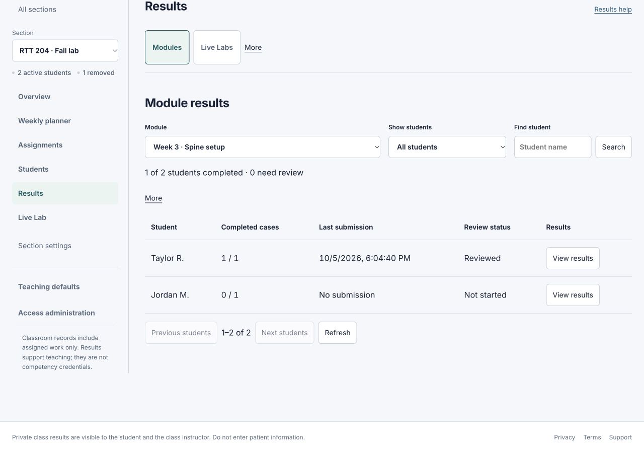
Six original browser screenshots, captured October 5, 2026, accompany the film: CBCT registration in three planes, Separated treatment-head teaching model, MLC leaf and drive mechanism, Separated kV detector layers, Plan a teaching week, Review fictional learner results. The CBCT capture uses the prostate planning-derived display attributed above (CC BY 4.0). Machine images show representative teaching geometry. The Weekly planner and results use entirely fictional class and learner records. No patient upload, private learner record or account detail appears. The original screenshot bytes are unchanged.
Previous full tour (v9)
The previous 2:24 film, poster and scene text remain available. Its original attribution and capture description follow.
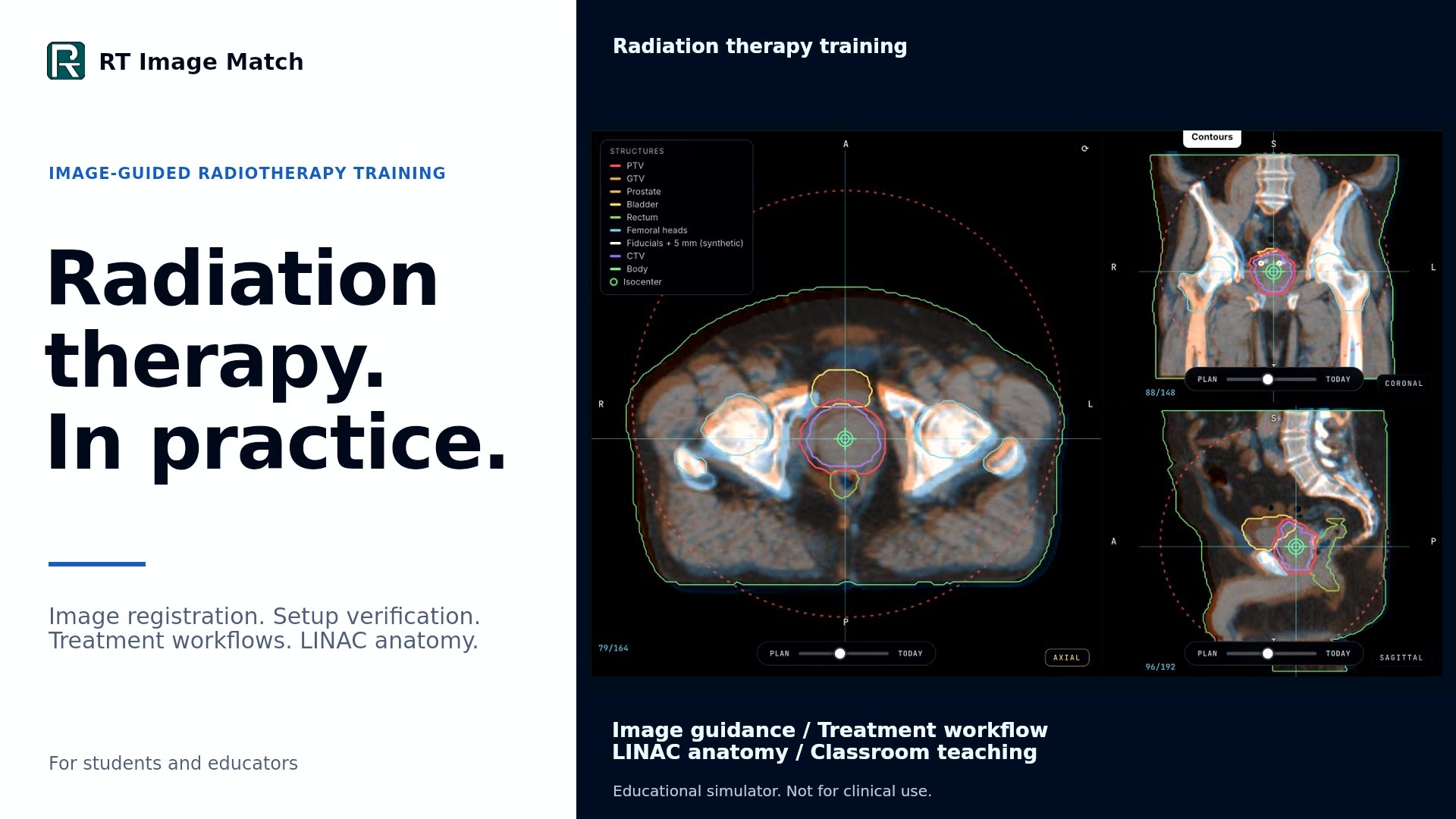
The retained 2 minute 24 second film shows image registration, a prostate treatment workflow, DIBH coaching preparation, LINAC anatomy, the case library and Classroom teaching tools. September 30 fictional-account recordings are combined with October 5 live-trainer and fictional Classroom captures. Seven fresh motion sequences keep image adjustments, slice review and machine orbits steady. Every scene preserves consecutive encoded frames at 25 fps, with brief reading holds and natural UI pauses; sequential browser capture is time-compressed and is not a claim of real-time 25 fps acquisition.
Native Higgsedit titles and framing sit outside the recorded software views. No source anatomy, displayed values or intermediate movement is generated, redrawn or interpolated. Independent matching is ungraded practice. Full Treatment shows the beginning of a VMAT arc, not a completed fraction. DIBH uses a local synthetic transcript fixture for coaching preparation; voice never starts imaging or delivery. Classroom names, schedules, attendance and results are fictional. The equipment uses representative teaching geometry, not manufacturer CAD, and has no clinical dose model. No source head or facial imaging, private learner records, account details or microphone recordings appear. One 1920 × 1080 landscape MP4 serves every screen. Educational use only; not for clinical decisions.
Prostate CBCT and Full Treatment: Thompson, Reid F., et al. (2023), Prostate-Anatomical-Edge-Cases, Version 1 (TCIA), CC BY 4.0. Planning-derived educational images use simulated daily transforms and overlays. Pelvis 2D/2D: Zuley, Margarita L., et al. (2016), TCGA-PRAD, Version 4 (TCIA), CC BY 3.0. Planning DRRs and simulated setup images are trimmed and proportionally scaled. The results shown here are in whole or part based upon data generated by the TCGA Research Network: cancer.gov/tcga.
Partial Breast L reference: Armato III, Samuel G., et al. (2015), LIDC-IDRI, Version 4 (TCIA), CC BY 3.0. The film frames the simulated room and respiratory monitor. Full collection citations and TCIA usage restrictions apply.
Music: “Inspired” by Kevin MacLeod (incompetech.com), licensed under Creative Commons Attribution 4.0. The excerpt runs from source time 32 to 176 seconds, with loudness normalization and entrance/ending fades; speed and pitch are unchanged. All capture audio is discarded. No speech, trainer sounds or microphone recordings are included. Typography uses Inter under the SIL Open Font License. No source author or organization is implied to endorse the film.
Previous 60-second film (v8)
The previous film, poster and scene text remain available for existing links. Their original credits follow.
The retained 60-second advertisement shows actual changes in the live trainer: planar and CBCT image blending, manual corrections, slice browsing, gantry rotation, simulated arc movement and a camera orbit. Recorded on October 5, 2026 using ordinary interface controls. Sequential browser frames preserve source order, with timing compressed for the edit; unchanged blend-control endpoints are removed. The closing camera orbit plays in reverse. Native Higgsedit titles and framing sit over the recorded footage. Source anatomy and displayed values are not generated or redrawn.
Matching uses untimed, ungraded practice. Machine Room uses a synthetic five-fiducial cube phantom; its geometric arc demonstration has no clinical dose model. No source head or facial imagery, private learner records or account details appear. One 1920 × 1080 landscape MP4 serves every screen. Educational use only; not for clinical decisions.
Prostate CBCT: Thompson, Reid F., et al. (2023), Prostate-Anatomical-Edge-Cases, Version 1 (TCIA), CC BY 4.0. The film captures planning-derived educational images with simulated daily transforms and overlays. Pelvis 2D/2D: Zuley, Margarita L., et al. (2016), TCGA-PRAD, Version 4 (TCIA), CC BY 3.0. Planning DRRs and simulated setup images are captured, trimmed and proportionally scaled. The results shown here are in whole or part based upon data generated by the TCGA Research Network: cancer.gov/tcga. Full collection citations and TCIA usage restrictions apply.
Music: “Inspired” by Kevin MacLeod (incompetech.com), licensed under Creative Commons Attribution 4.0. The 60-second excerpt starts at source time 48 seconds, with loudness normalization and entrance/ending fades. No speech, trainer sounds or microphone recordings are included. Typography uses Inter under the SIL Open Font License. No source author or organization is implied to endorse the advertisement.
Previous two-minute film (v7)
The previous film and its poster remain available for existing links. Their original credits follow.
The two-minute video follows one prostate Full Treatment case through plan review, CBCT acquisition and matching, couch correction, kV fiducial verification and the start of simulated VMAT delivery. It retains Partial Breast L · DIBH IMRT preparation, then shows Machine Room gantry movement, treatment-head components, the MLC leaf study and kV detector layers. Classroom recordings show the Weekly planner, assignments, Live Lab teaching and module results. One landscape edit serves every screen.
Native Higgsedit graphics add task descriptions and a closing Launch trainer message outside the footage. Filmed actions retain their wall-clock speed, fixed crops and eight-frame editorial dissolves. Partial-breast room, coaching and trace details are synchronized crops from one recording. No source anatomy, result values or motion are generated or interpolated.
Music: “Inspired” by Kevin MacLeod (incompetech.com), licensed under Creative Commons Attribution 4.0. The video uses seconds 32–152, with entrance/ending fades and loudness normalization; speed and pitch are unchanged. All capture audio is discarded. The soundtrack contains no speech, trainer sounds or microphone recordings. These edits do not imply endorsement by the composer.
Captured on September 30, 2026 in local fictional-account fixtures. The prostate fraction starts with an authored setup error. Manual image drags, visible-review-guided refinement, held couch correction, kV verification and Beam On use the actual simulator controls and grading. Selected preparation, kV acquisition waits and refinement are omitted. The delivery excerpt shows the start of an arc, not a completed fraction. The DIBH scene uses a local synthetic audio/transcript fixture through held push-to-talk; it shows preparation only, and voice never starts imaging or treatment. Classroom names, schedules, attendance and results are fictional. The case picker is text-only; no head-derived MR image is published.
The prostate treatment, Machine Room patient and Live Lab demonstration use Prostate-Anatomical-Edge-Cases (TCIA), licensed CC BY 4.0. The Partial Breast L exercise’s reference source is LIDC-IDRI (Armato et al., TCIA), licensed CC BY 3.0; this film frames the room and breathing monitor. Full collection citations and TCIA usage restrictions apply. Equipment internals are representative teaching models, not manufacturer CAD.
Retained CBCT recording and captured images
The September 30 playback repair corrects the CBCT videos’ timing and compatibility metadata. Every recorded frame and the complete narration are preserved without re-encoding. The former landing-page example images remain available with their original pixels.
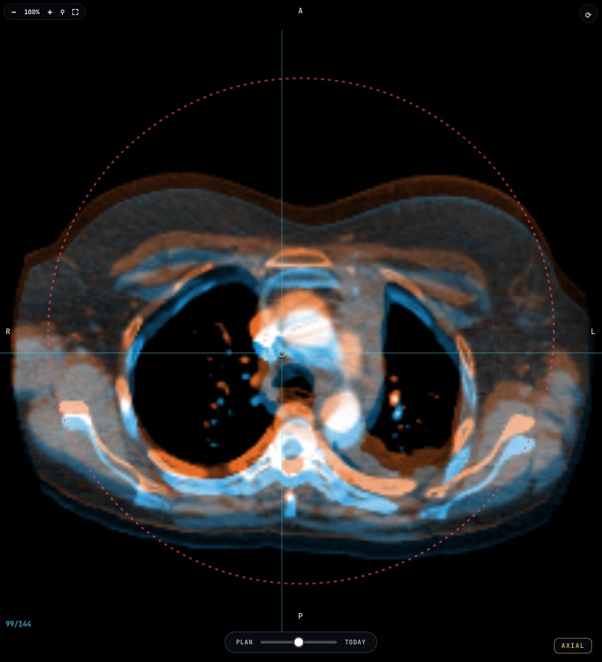
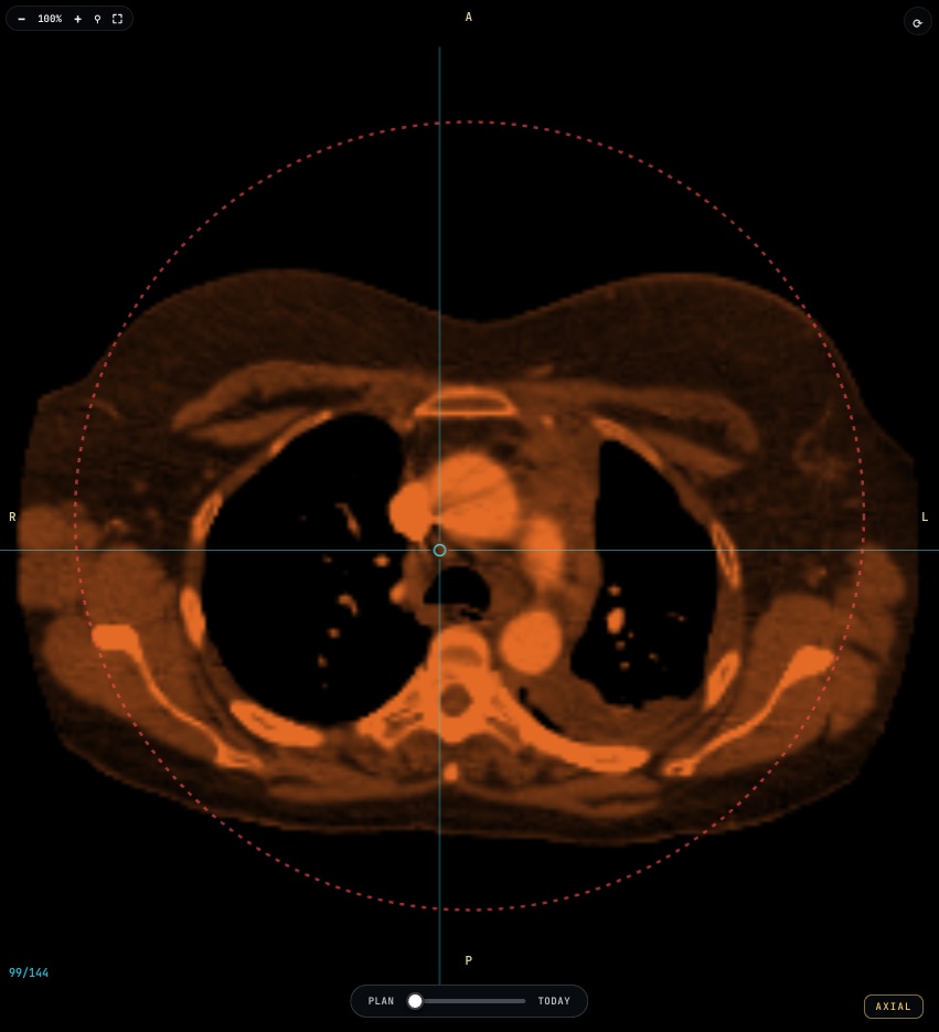
The retained narrated source recording follows one complete Mediastinal Lymph Nodes registration exercise. The 75-second edit comes from a continuous trainer recording, with real pointer drags, rotation, keyboard adjustments, blend changes, slice review and two actual checks. Automated UI input in a local synthetic-account fixture operates the ordinary controls; refinement uses only the visible first report. No Auto Match or hidden-answer access is used. Editing removes idle pauses, crops the same frames for phones, adds simple framing and cursor emphasis, and aligns conversational AI narration (Cillian, generated with Higgsfield) at its original speed and pitch. Source anatomy and captured report values were not generated or changed by the edit. The account header is excluded, and enlarged reports omit attempt history, elapsed-time and personal-best fields. Any remaining timing or status belongs only to the synthetic-account fixture. The landing-page interactive example uses six lossless plane crops from this same recording: one before and one after refinement for each of the axial, coronal and sagittal views. Both states retain the same slice, framing and 50% blend. The Before/After control switches captured states; it does not calculate a registration or score.
The source CT and node annotations are LNQ case_0412 from Mediastinal Lymph Node Quantification (TCIA), licensed CC BY 4.0. The public field is thoracic; it does not include head or facial anatomy. See the full collection citation below. Source licensing does not establish research-participant consent or other source-specific privacy facts.
The optional 12-second silent Full Treatment tour is edited from the previously recorded prostate Full Treatment sequence. The 24-second Machine Room edit uses the recorded training linac. They add chapter titles, crops, timing changes and inspection pauses. Phone versions reframe the same reviewed imagery. No generated anatomy was added. The prostate source is Prostate-MRI-US-Biopsy (TCIA), licensed CC BY 4.0. Equipment geometry is an educational representation, not manufacturer CAD.
Captured before and after images
- Axial: before refinement · after refinement (original PNGs: before · after)
- Coronal: before refinement · after refinement (original PNGs: before · after)
- Sagittal: before refinement · after refinement (original PNGs: before · after)
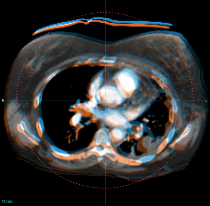
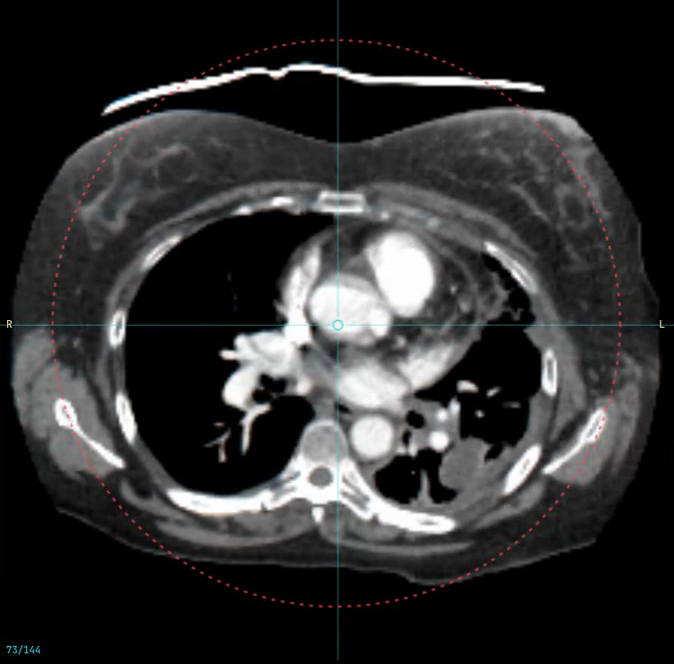
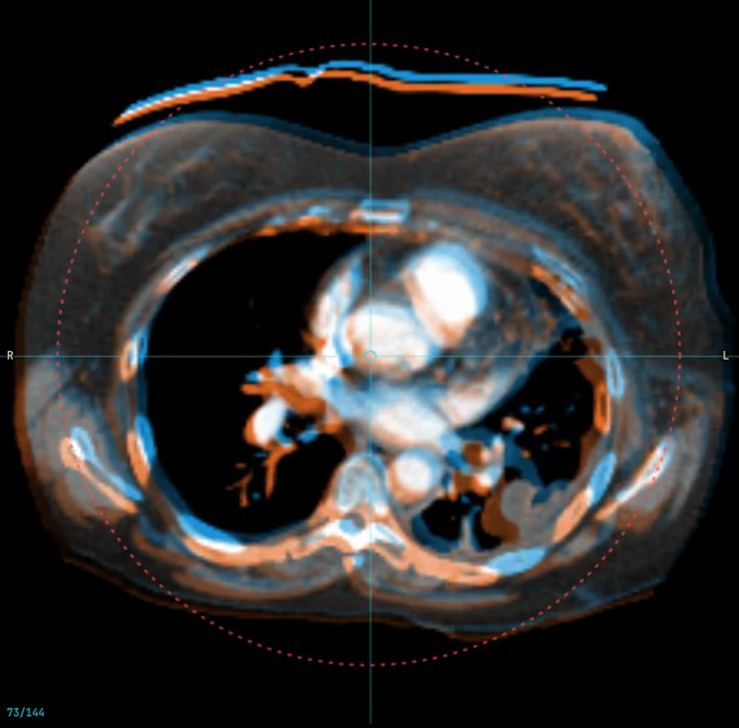
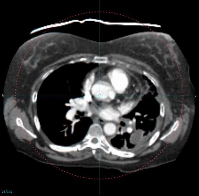
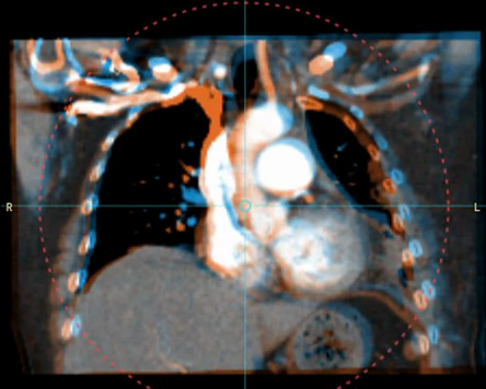
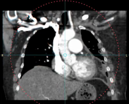
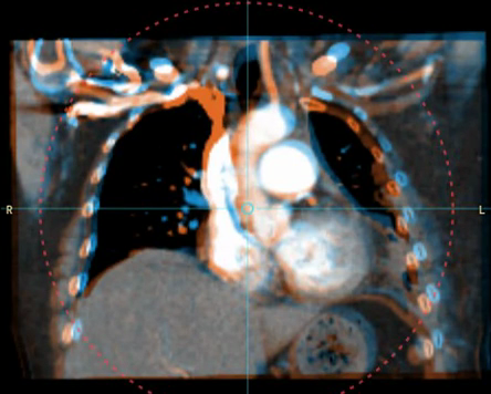
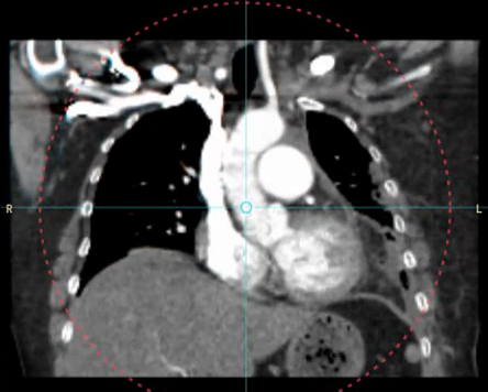
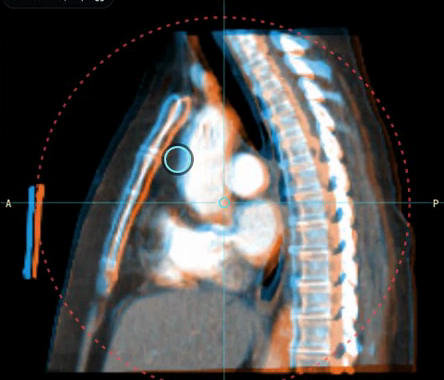
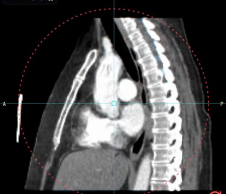
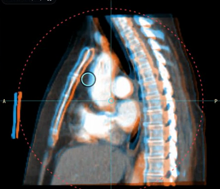
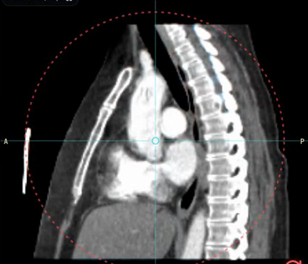
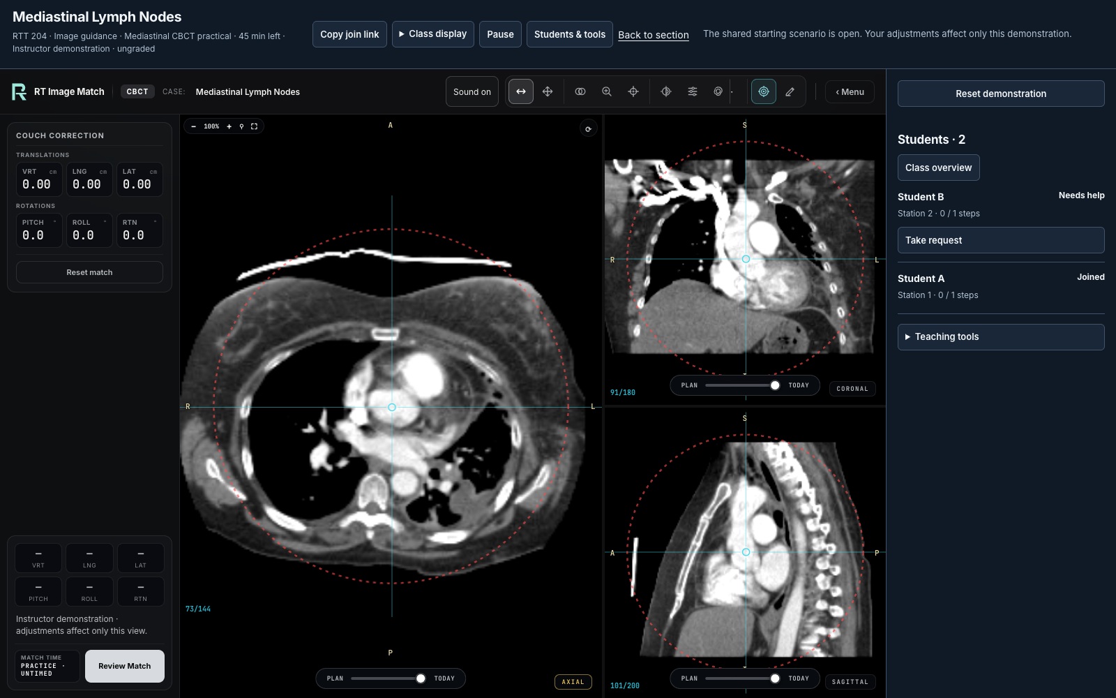
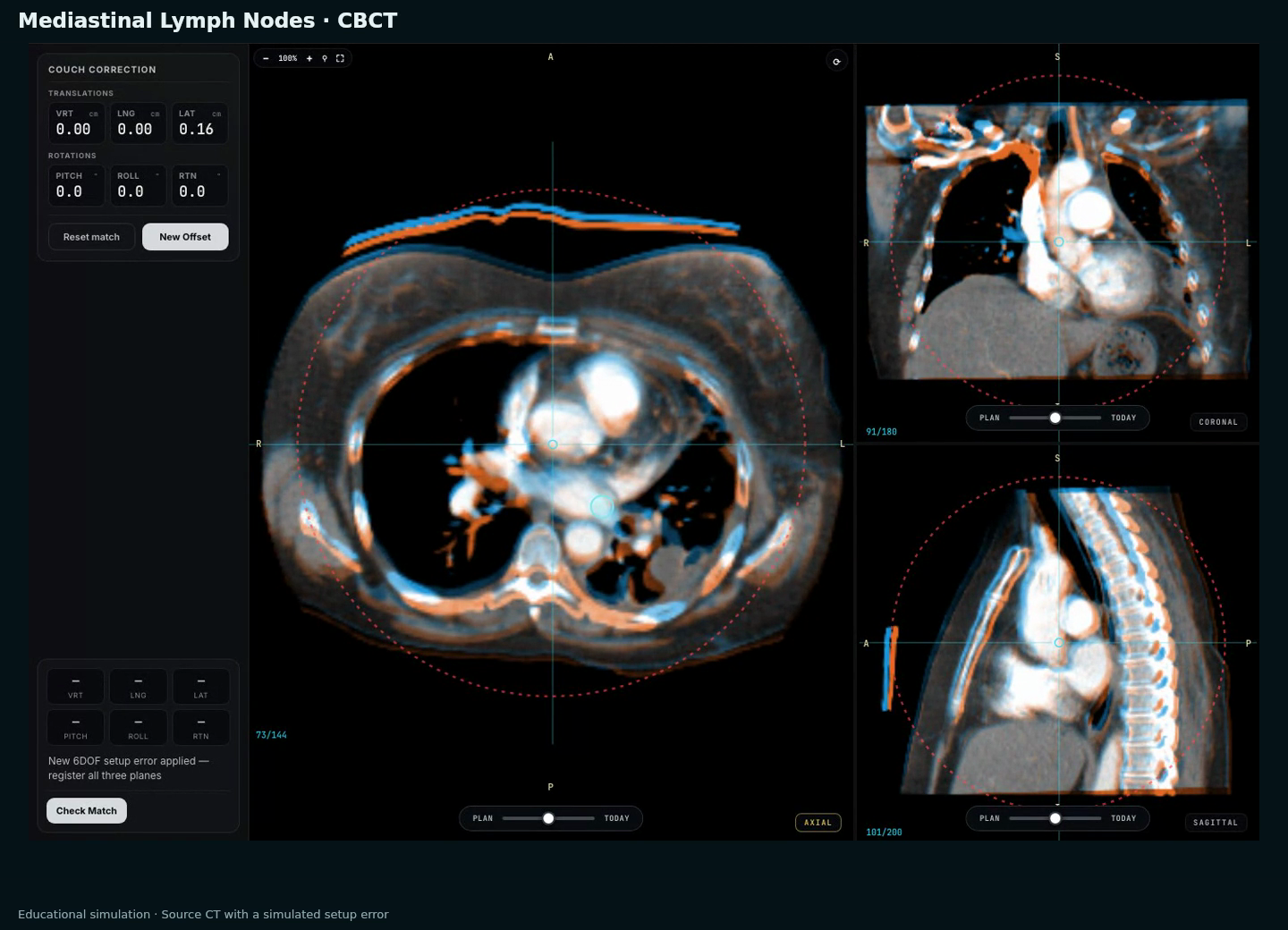
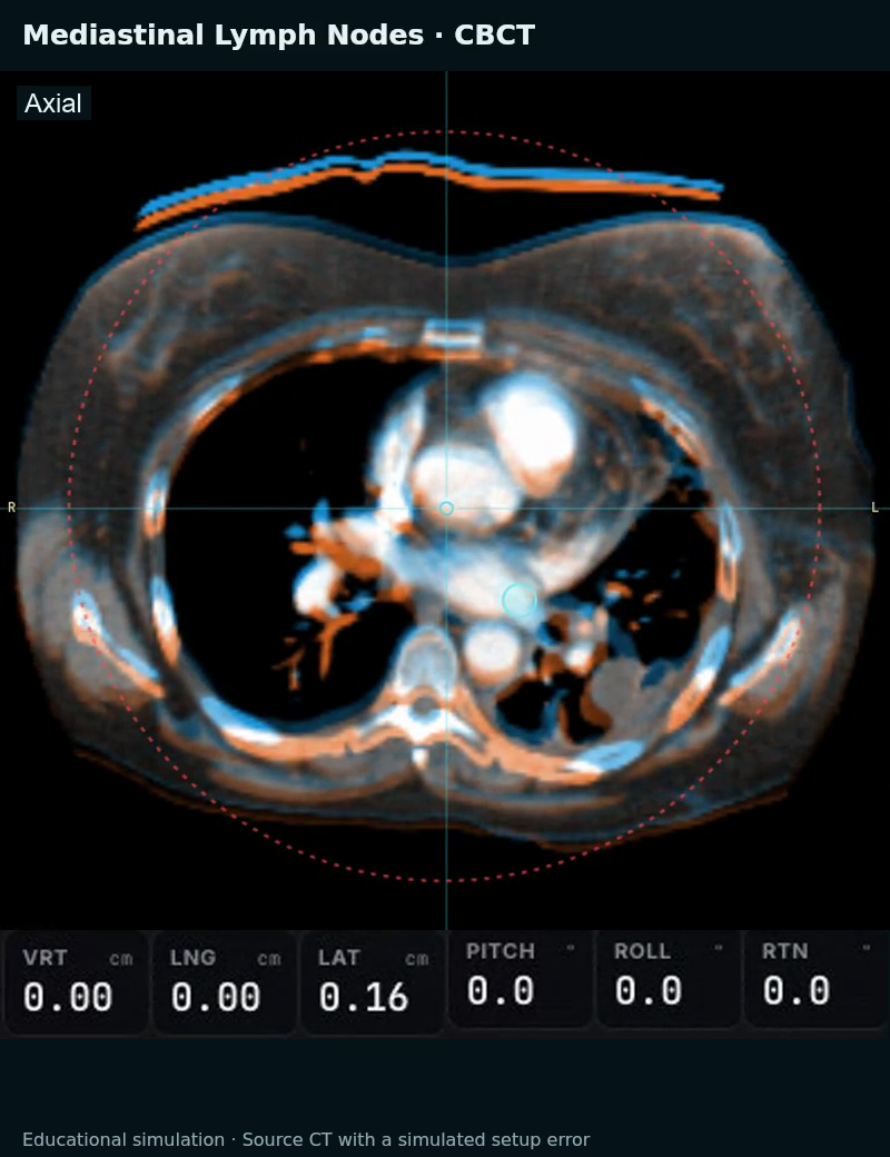
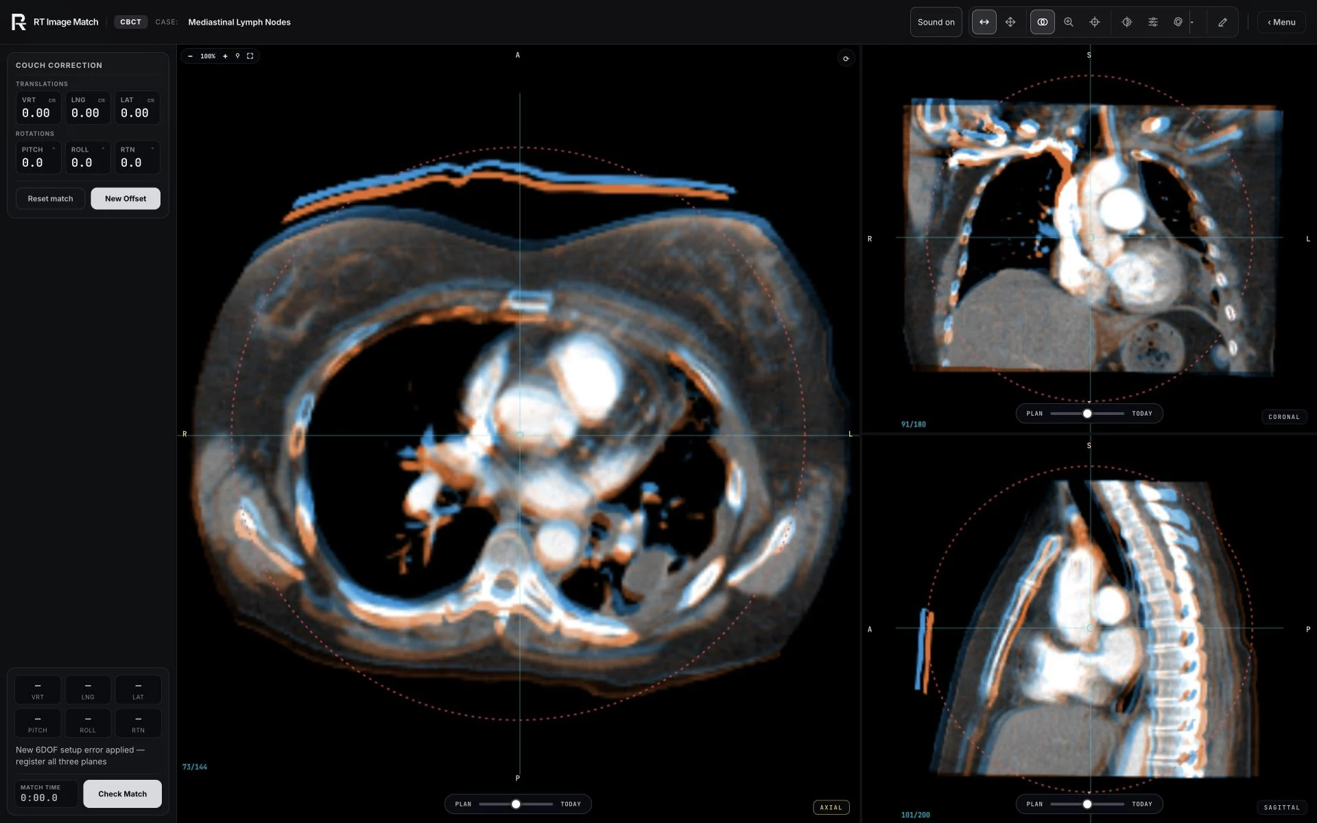
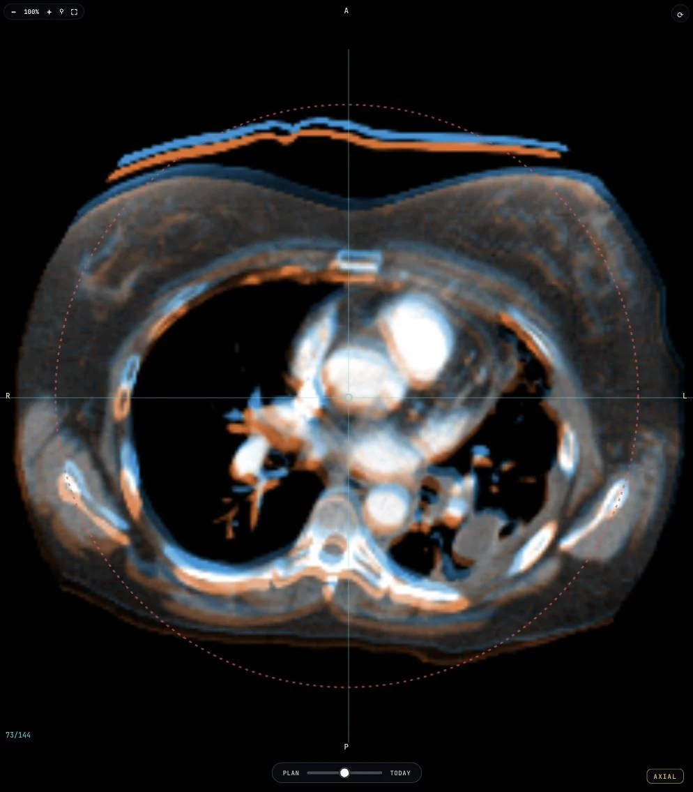
The lossless WebP previews preserve every decoded RGB pixel of the original PNG crops. Crops are from 39 and 73 seconds of the reviewed walkthrough. Axial slice 73/144, coronal 91/180 and sagittal 101/200 are unchanged between the two states. No generated anatomy, image interpolation or extra correction is applied to these captures.
Retained CBCT source files
The previous CBCT recording includes historical AI narration. The recording remains available for provenance; the separate landing-page CBCT example has been removed: desktop recording · phone recording · desktop poster · phone poster.
What the images and results mean
For the CBCT exercise, source CT serves as the planning reference. Today is derived from the same CT with a simulated rigid setup error; it is not a separately acquired daily CBCT. Contour overlays are off in the walkthrough. When enabled in the trainer, yellow contours identify source node annotations. The 5 mm and 10 mm expansions from those annotations are geometric teaching aids, not clinical target volumes or prescribed margins. Six degrees of freedom means three translations and three rotations.
The feedback table uses the actual first check: 7.7 mm combined translation residual, with lateral and vertical outside this exercise’s limits. The final check displays a 0.3 mm combined translation residual, with all three translations and three rotations within their separate limits (±3 mm per translation axis; ±2° per rotation axis). These are simulator scores, not measured scanner accuracy, clinical action thresholds or clinical approval.
Terminology references: DICOM spatial registration and AAPM Task Group 132.
Classroom previews
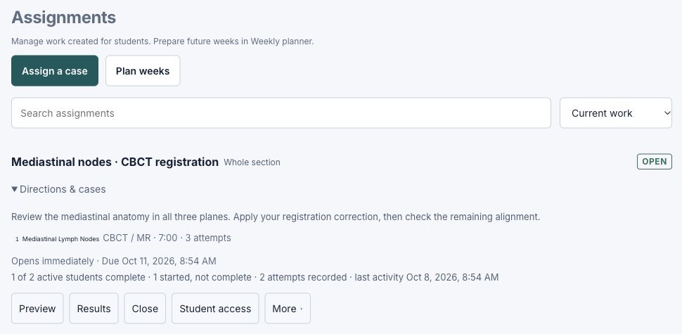
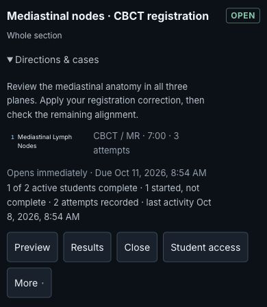
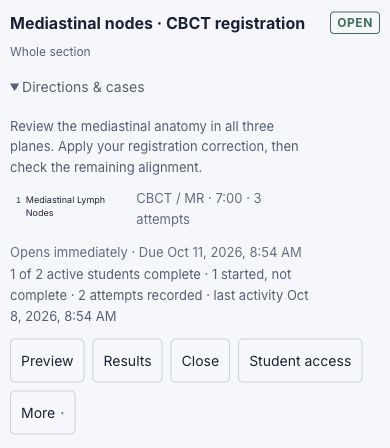
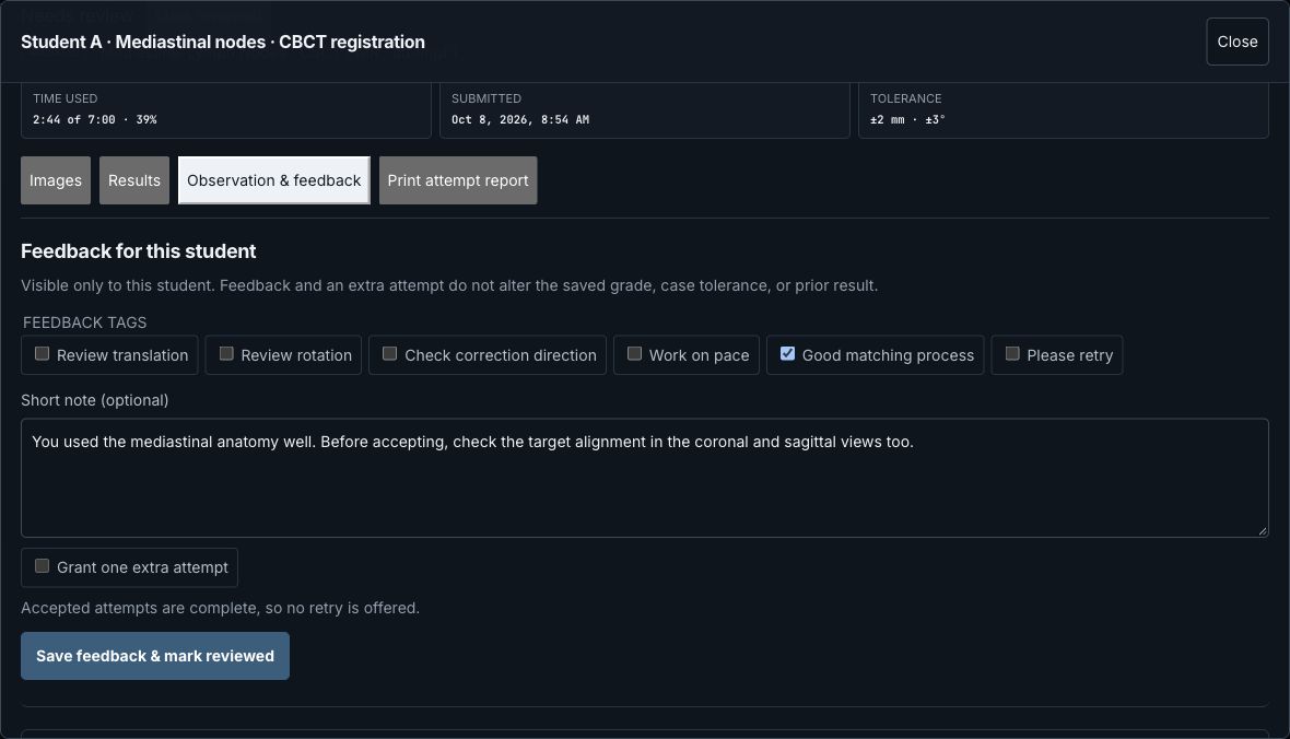
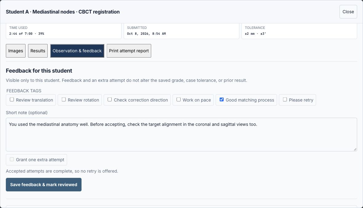
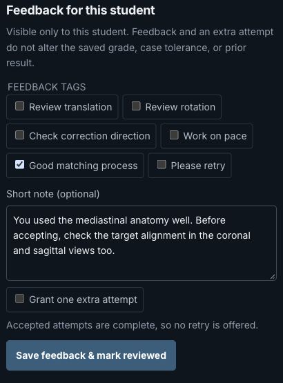
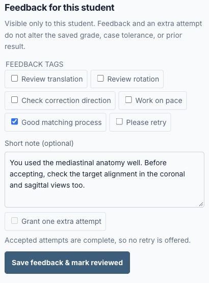
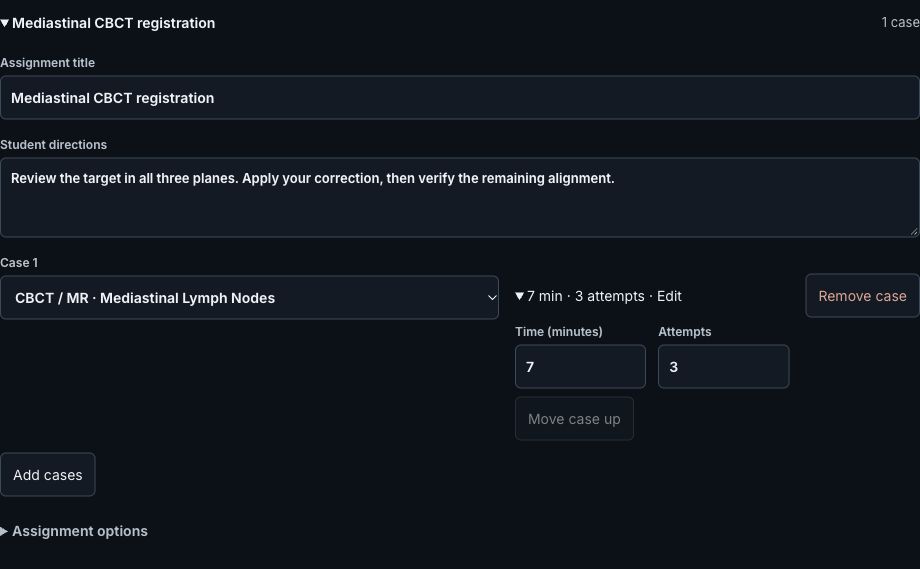
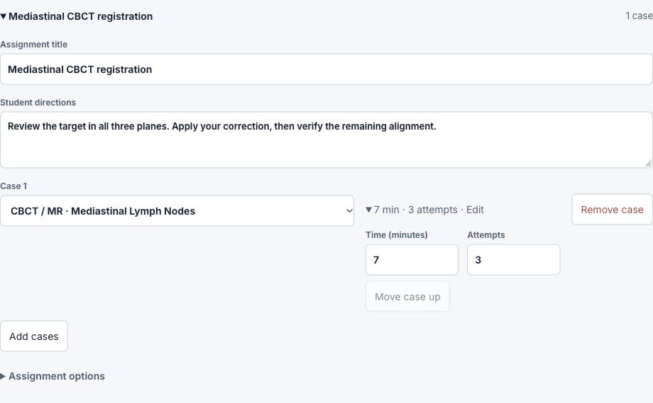
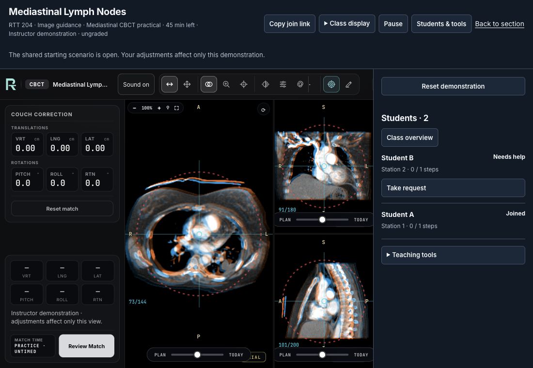
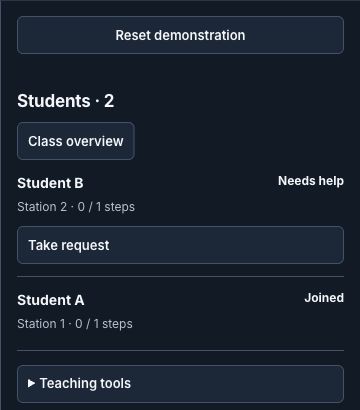
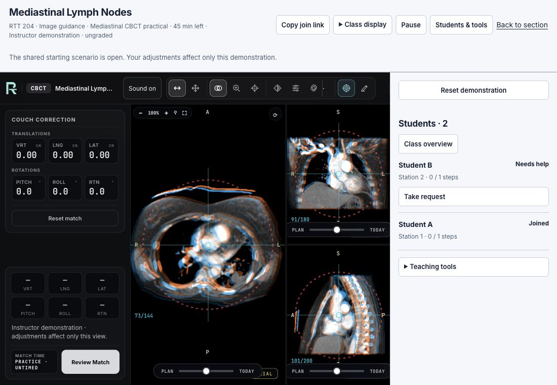
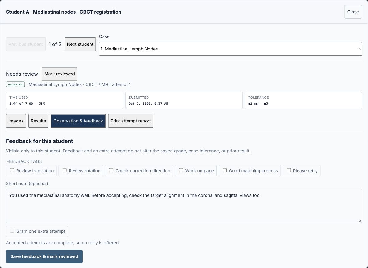
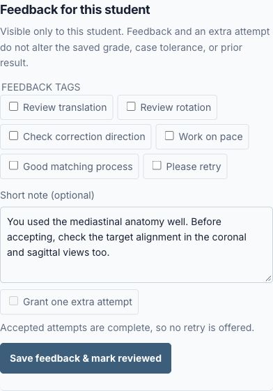
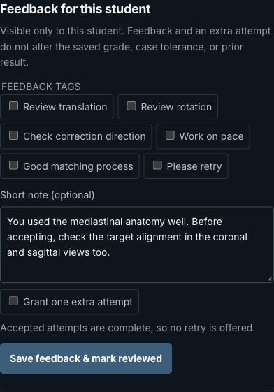
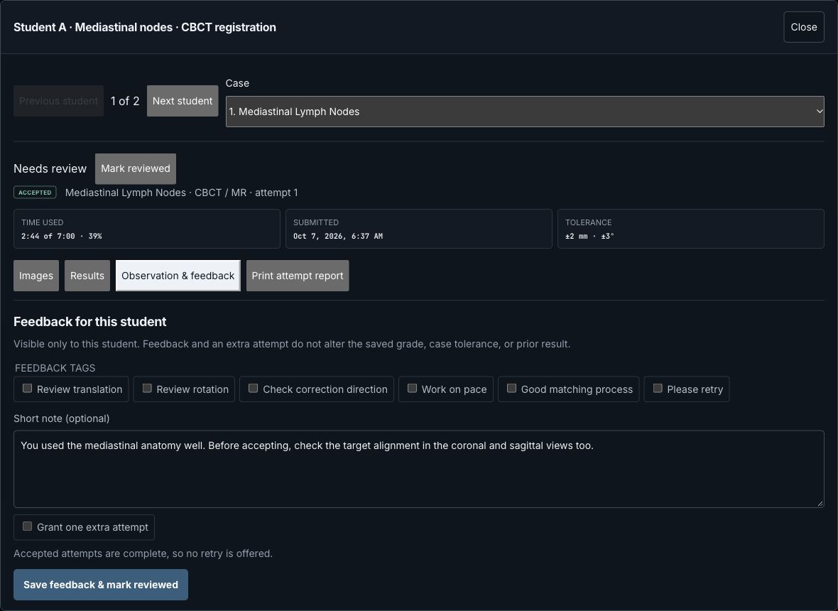
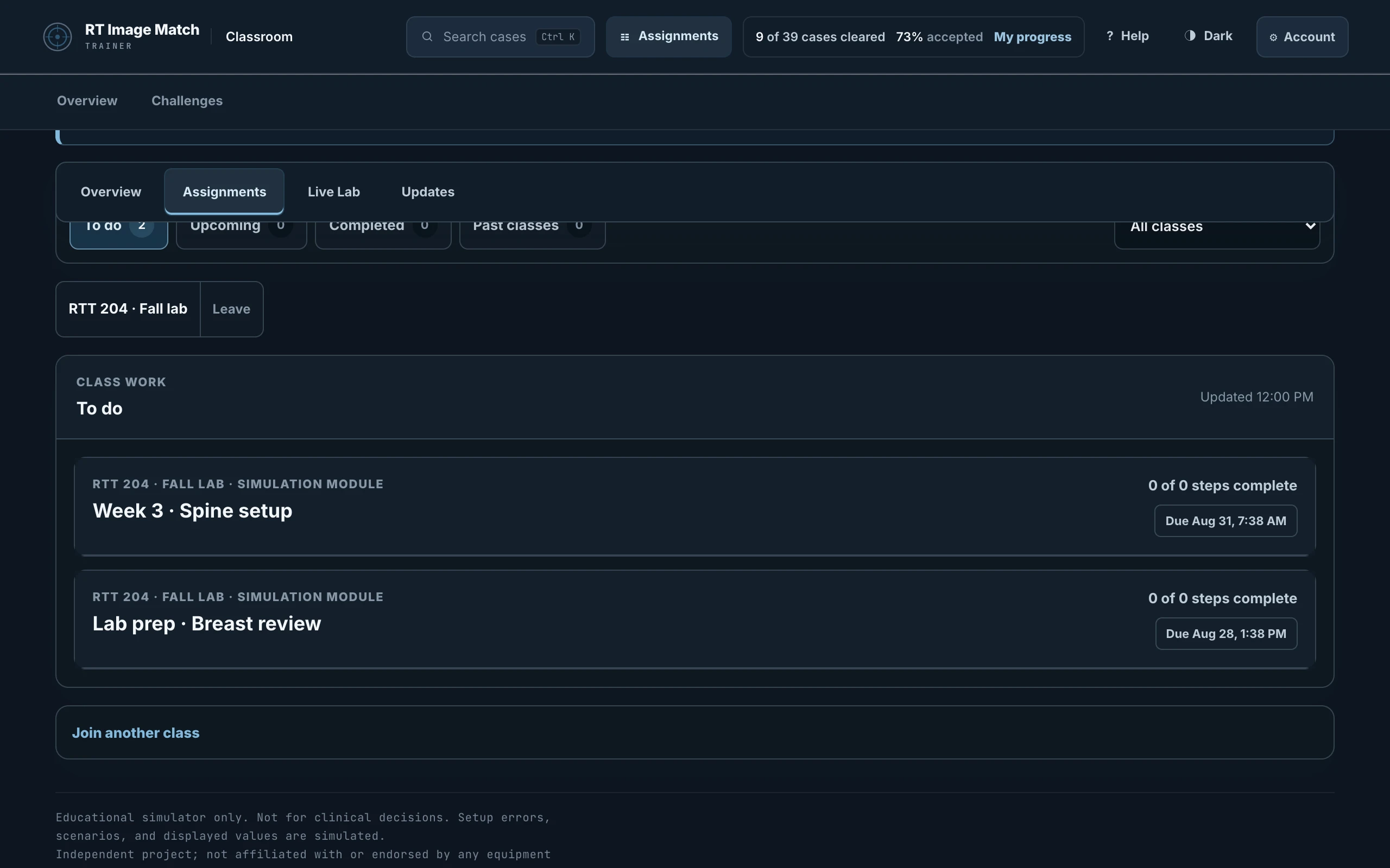
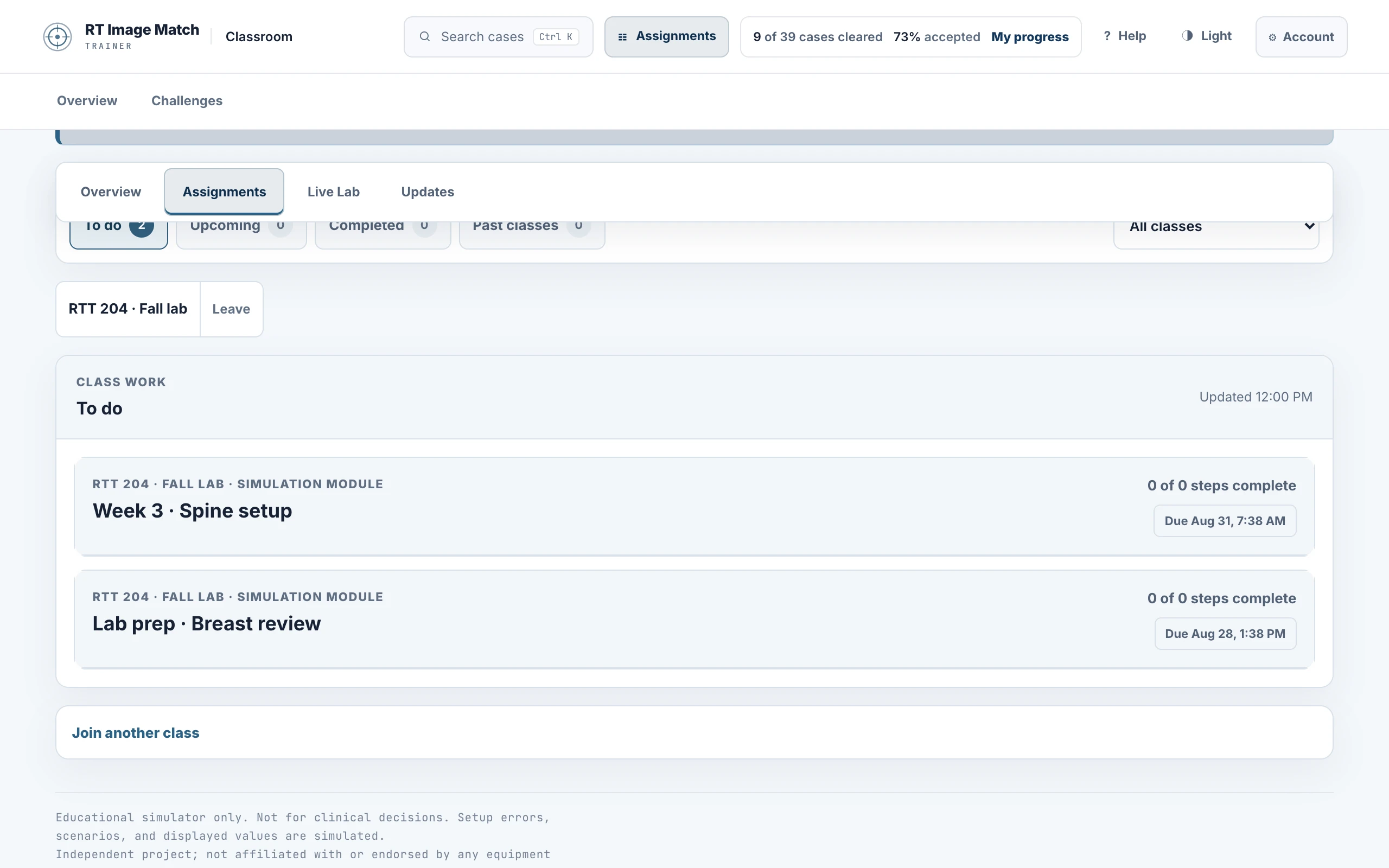
The current Classroom walkthrough shows Assign practice, Teach live, and Review & feedback. Eight October 8, 2026 captures show a published assignment and individual feedback; four October 7 captures show Live Lab. Separate desktop and phone layouts are available in both appearances. Phone images focus on assignment details, the student help roster, and the feedback note; enlargement opens the desktop task. Class details, dates, students, results and feedback are fictional; no real account or student record appears. The examples do not represent a recorded learner journey.
The desktop Live Lab images show thoracic anatomy from LNQ case_0412 in Mediastinal Lymph Node Quantification (TCIA), licensed CC BY 4.0. Full collection citation appears below. Plan and Today use the same source CT with a fictional rigid setup error, viewed with 50% color wash; Today is not a separately acquired daily CBCT. No source anatomy is generated or edited, and no head, facial anatomy or direct identifier is visible. Modules, feedback and phone Live Lab crops contain no anatomical images.
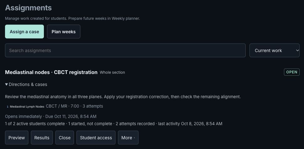
The October 8 Classroom refinement uses new native screenshots of a published assignment and individual feedback in desktop and phone layouts, in both appearances. All class details, student work and notes are fictional local fixture data. The feedback view shows an unsaved selection of “Good matching process.” Native JPEG crops are unchanged; no generated interface or anatomy is included. The October 7 Live Lab captures are retained.
October 8 Classroom screenshots
- classroom-refined-assign-desktop-dark.jpg
- classroom-refined-assign-desktop-light.jpg
- classroom-refined-assign-phone-dark.jpg
- classroom-refined-assign-phone-light.jpg
- classroom-refined-review-desktop-dark.jpg
- classroom-refined-review-desktop-light.jpg
- classroom-refined-review-phone-dark.jpg
- classroom-refined-review-phone-light.jpg
October 7 instructor images (Live Lab remains current)
- classroom-tools-modules-desktop-dark.jpg
- classroom-tools-modules-desktop-light.jpg
- classroom-tools-modules-phone-light.jpg
- classroom-tools-modules-phone-dark.jpg
- classroom-tools-live-desktop-dark.jpg
- classroom-tools-live-phone-dark.jpg
- classroom-tools-live-desktop-light.jpg
- classroom-tools-live-phone-light.jpg
- classroom-tools-review-desktop-light.jpg
- classroom-tools-review-phone-light.jpg
- classroom-tools-review-phone-dark.jpg
- classroom-tools-review-desktop-dark.jpg
Previous classroom walkthrough and student views
- classroom-instructor-live-desktop-dark.jpg
- classroom-instructor-live-desktop-light.jpg
- classroom-instructor-live-phone-dark.jpg
- classroom-instructor-live-phone-light.jpg
- classroom-walkthrough-assign-desktop-dark.jpg
- classroom-walkthrough-assign-desktop-light.jpg
- classroom-walkthrough-assign-phone-dark.jpg
- classroom-walkthrough-assign-phone-light.jpg
- classroom-walkthrough-practise-desktop-dark.jpg
- classroom-walkthrough-practise-desktop-light.jpg
- classroom-walkthrough-practise-phone-dark.jpg
- classroom-walkthrough-practise-phone-light.jpg
- classroom-walkthrough-review-desktop-dark.jpg
- classroom-walkthrough-review-desktop-light.jpg
- classroom-walkthrough-review-phone-dark.jpg
- classroom-walkthrough-review-phone-light.jpg
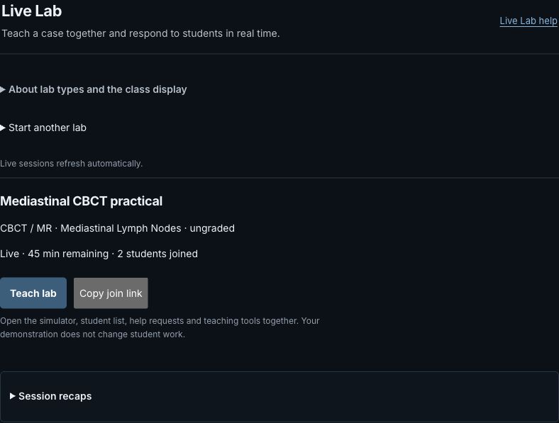
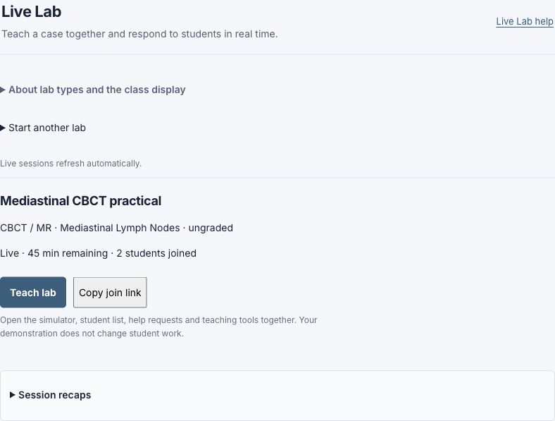
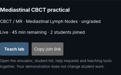
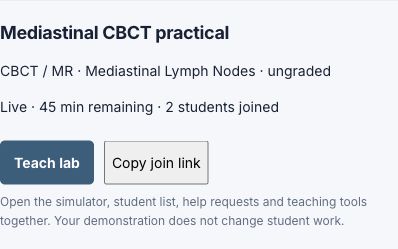
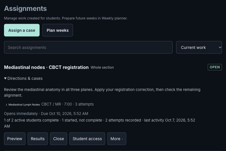
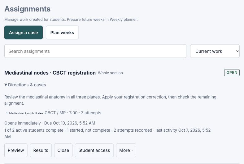
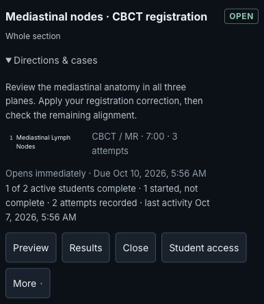
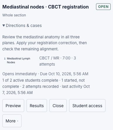
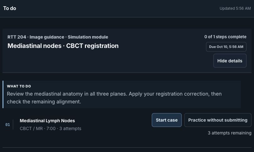
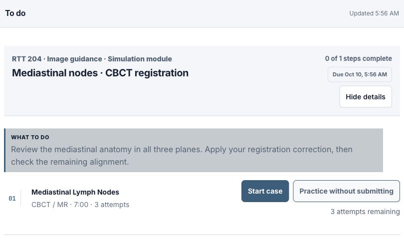
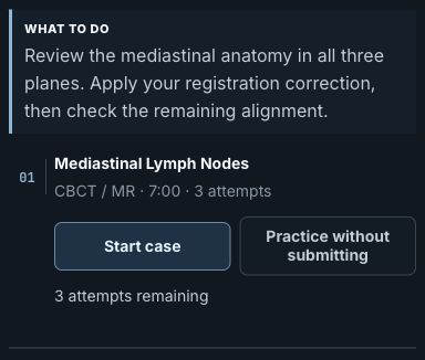
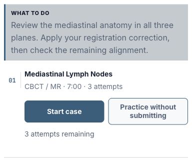
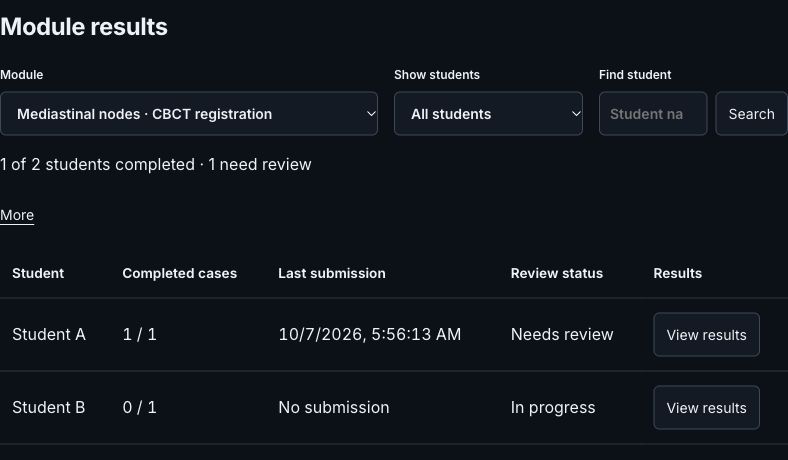
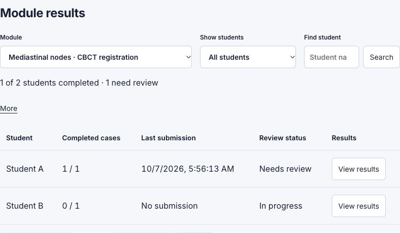
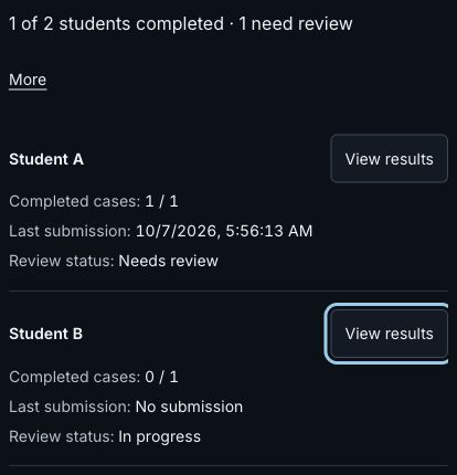
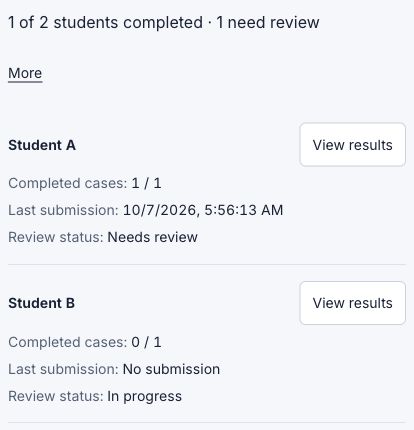
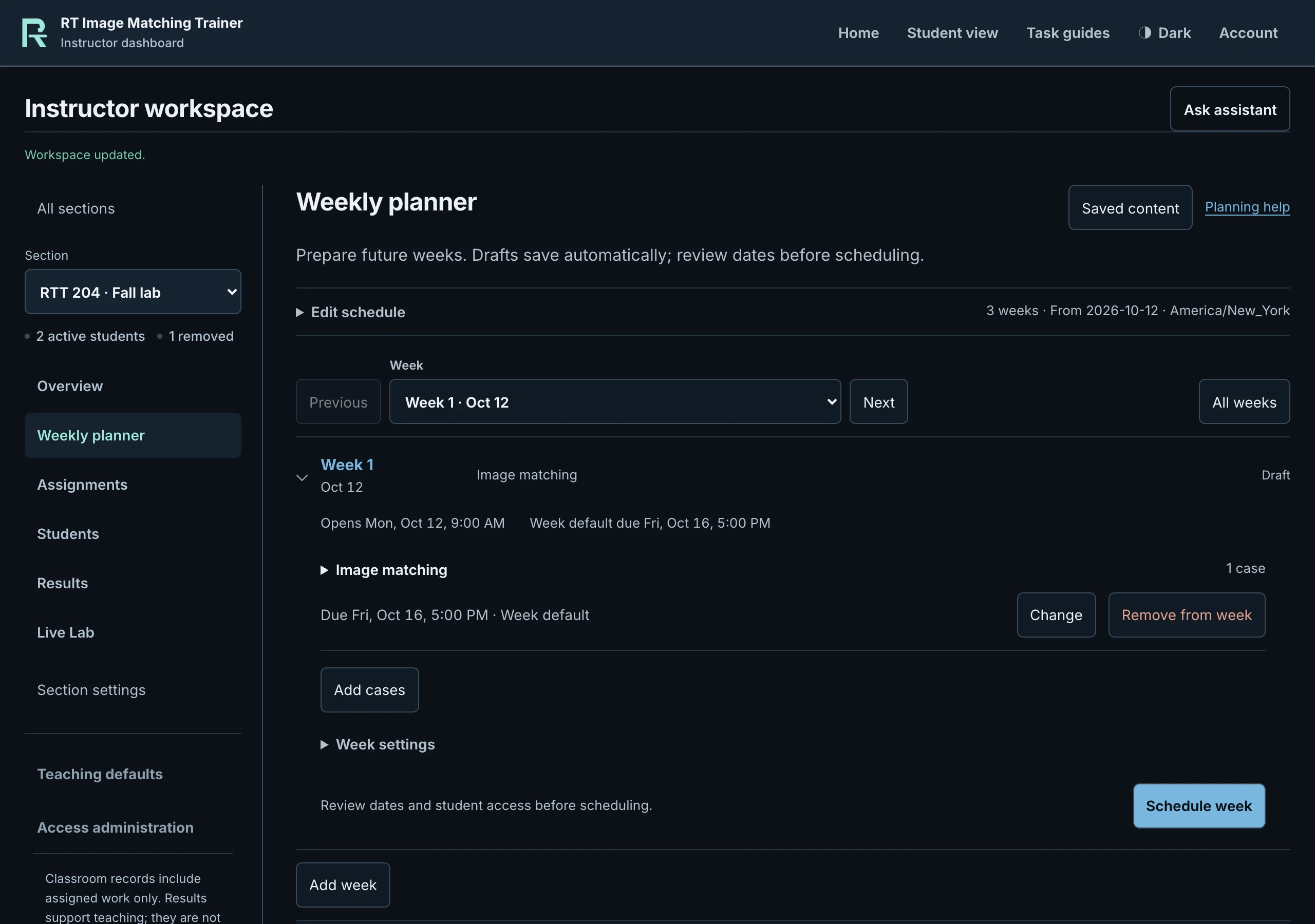
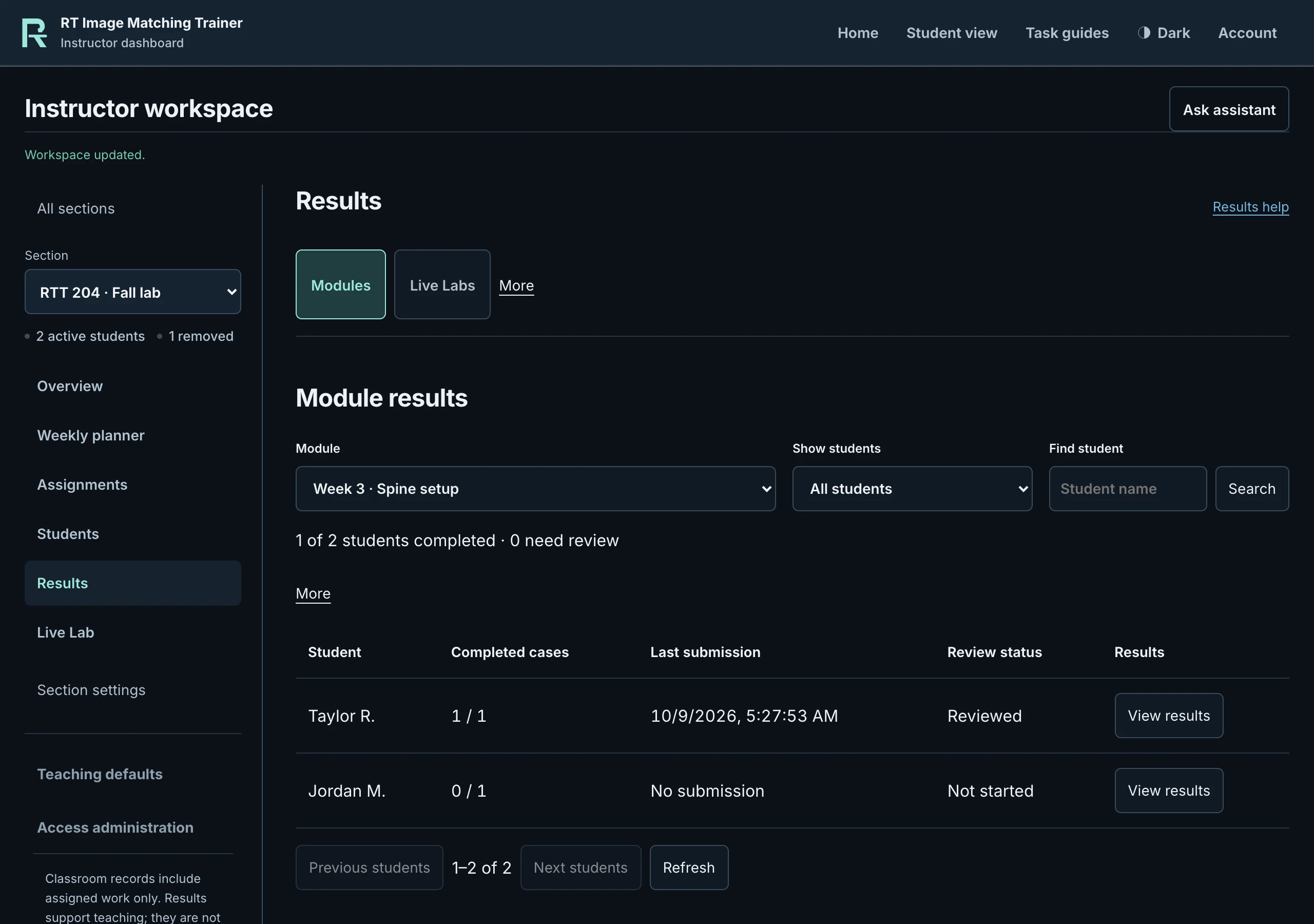
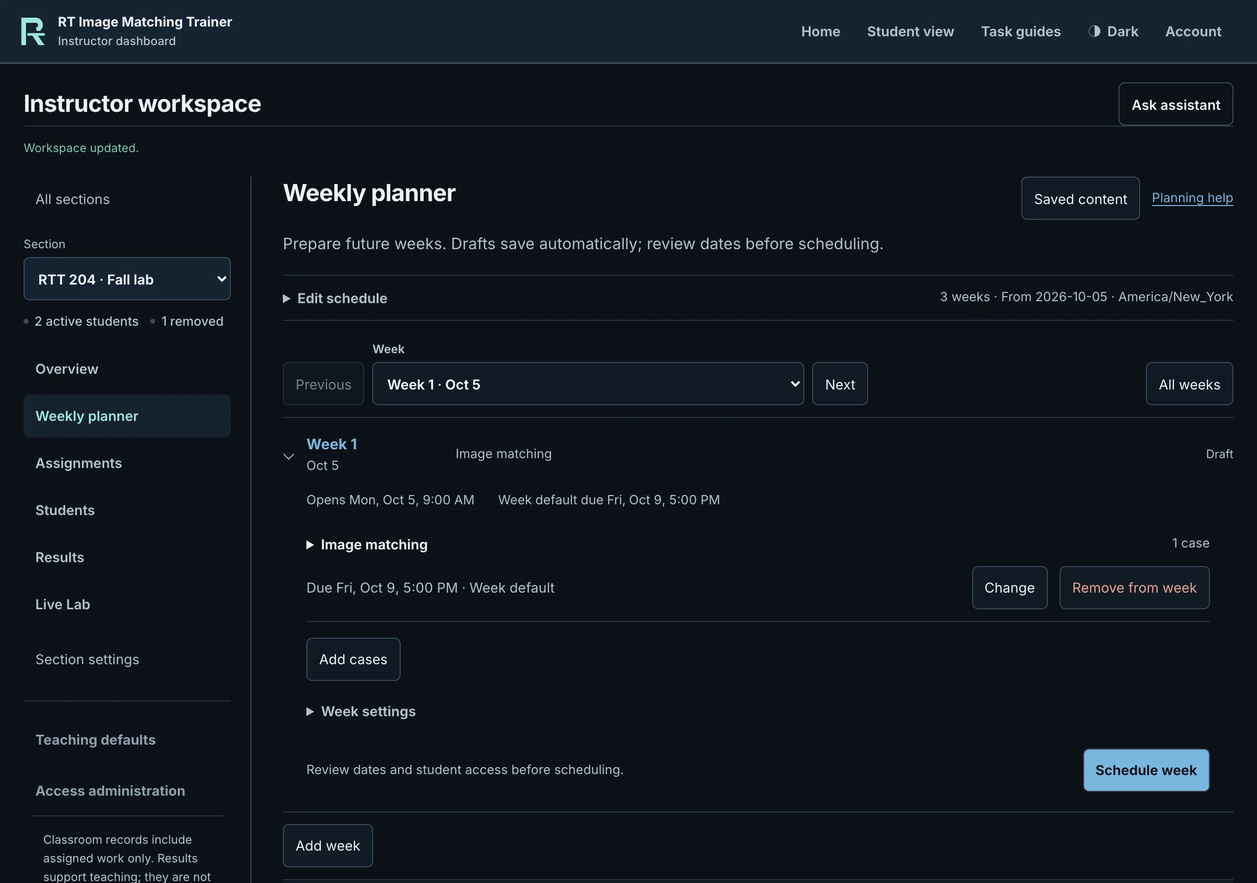
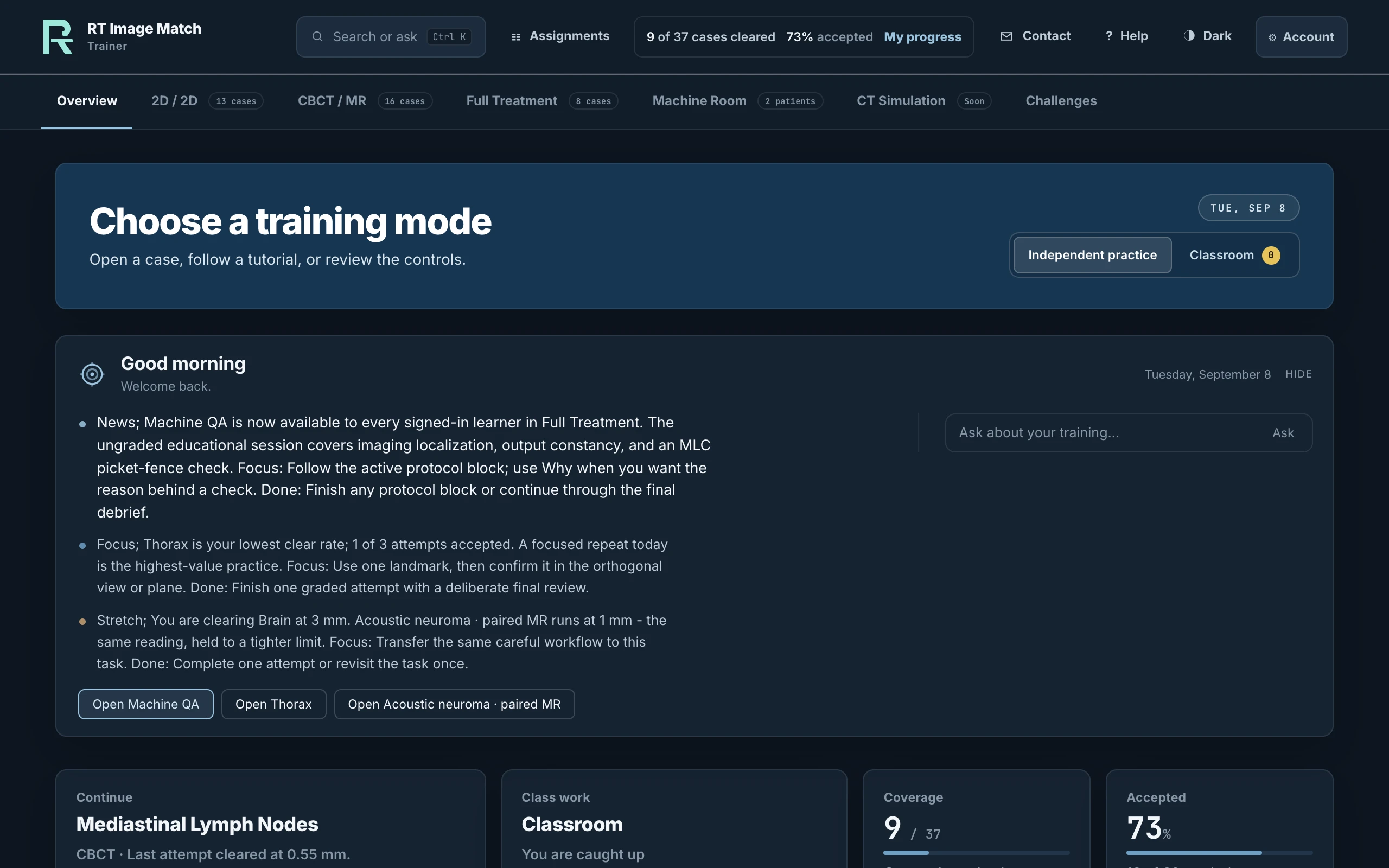
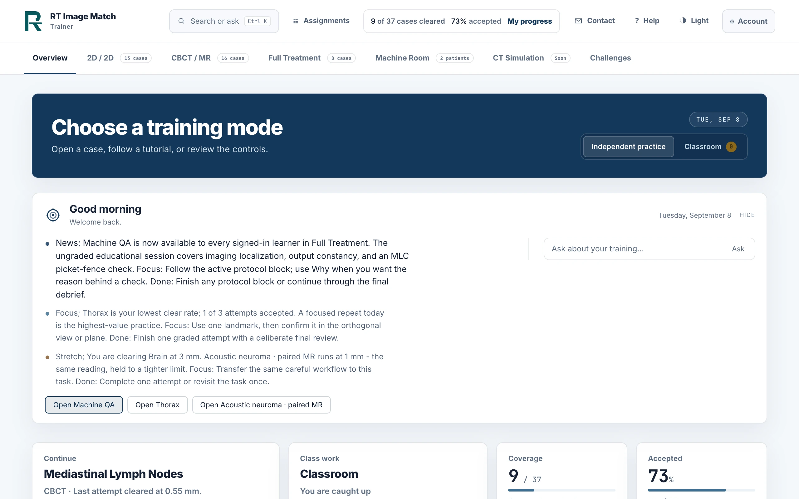
The previously captured Weekly planner, Live Lab overview and Results screenshots show the RT Image Match interface with local fictional class data. All names, teaching plans, attendance and results are invented. No production account, student record or patient image appears in these captures.
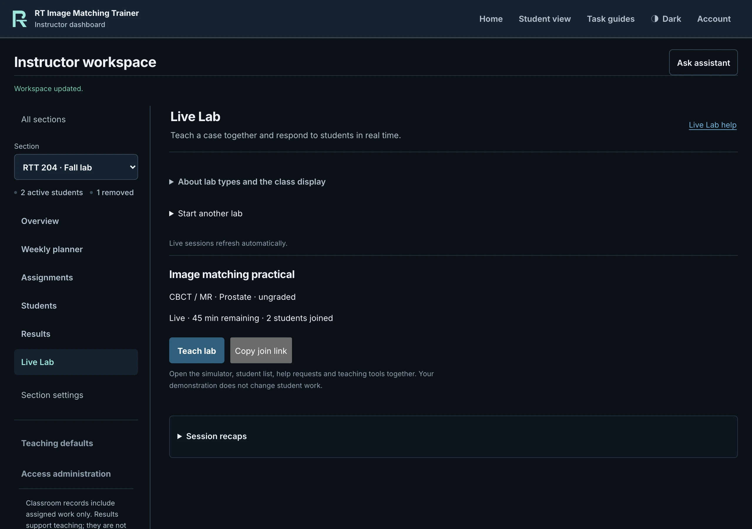
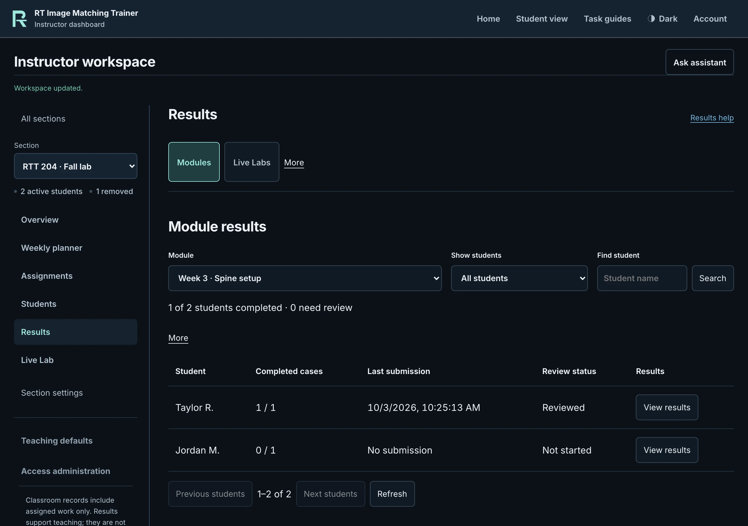
Original recording and supporting capture files
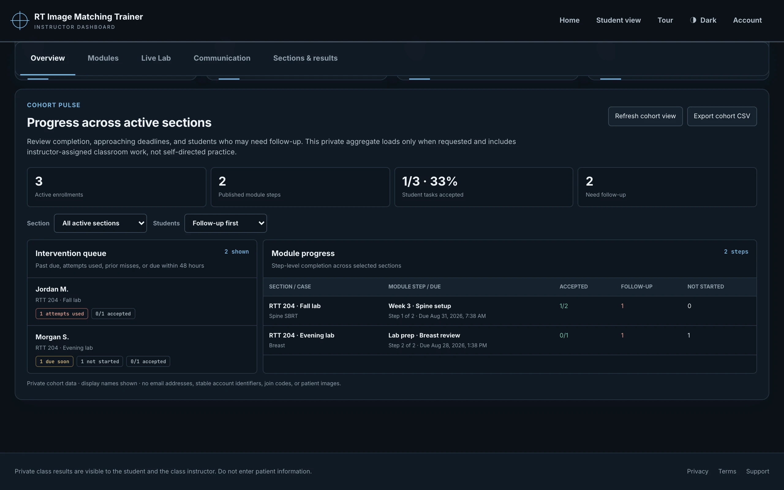
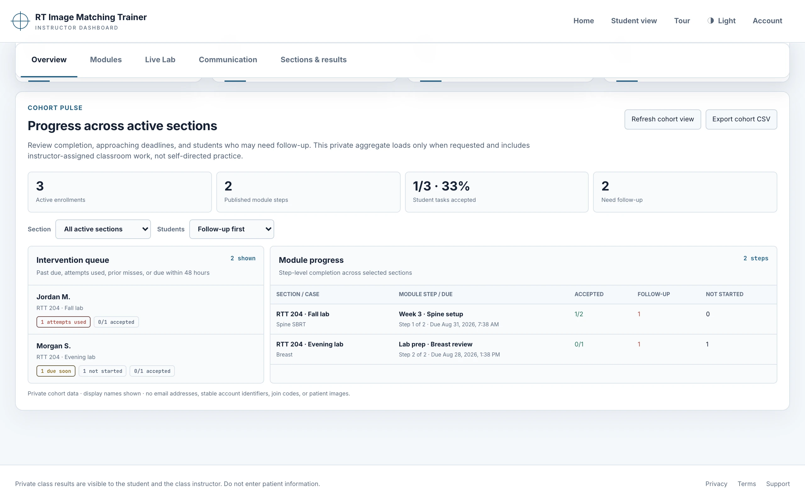
Previously reviewed source recordings and supporting captures remain available for attribution and comparison. Classroom and trainer-home examples use fictional learner records.
- classroom-instructor-dark.webp — previous instructor overview
- classroom-instructor-light.webp
- classroom-student-dark.webp
- classroom-student-light.webp
- trainer-home-dark.webp
- trainer-home-light.webp
- treatment-story.mp4
- treatment-compare.webp — original prostate matching preview
- treatment-compare-mobile.webp — phone version of the original prostate preview
- treatment-verification.webp
- treatment-delivery.webp
- machine-story.mp4
- machine-arms.webp
- machine-gantry.webp
- machine-internals.webp
- treatment-story-mobile.mp4
- machine-story-mobile.mp4
- treatment-verification-mobile.webp
- treatment-verified-mobile.webp
- treatment-delivery-mobile.webp
- machine-gantry-mobile.webp
- machine-arms-mobile.webp
- machine-internals-mobile.webp
- treatment-verified.webp
- machine-tour-mobile-poster-v2.png
- machine-tour-mobile-v2.mp4
- machine-tour-poster-v2.png
- machine-tour-v2.mp4
- scroll-tour-mobile-v2.mp4
- scroll-tour-v2.mp4
- feature-film-v10.mp4
- feature-film-v10-poster.jpg
Scroll-controlled matching and machine demonstration
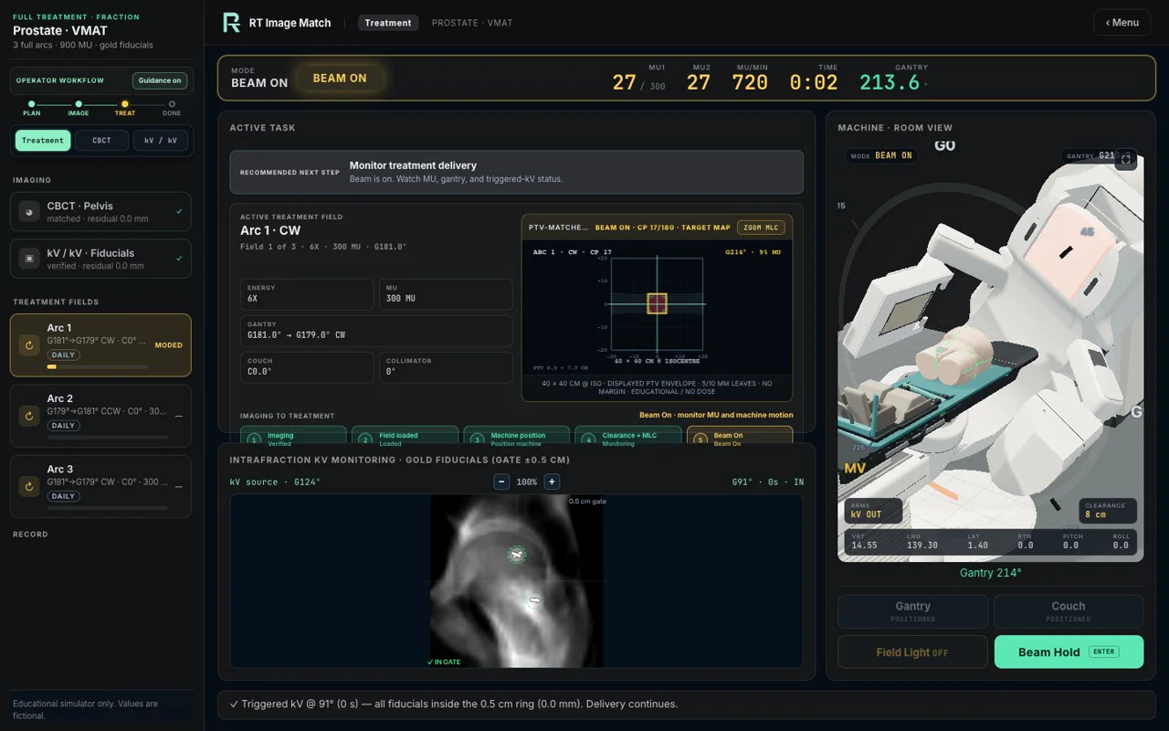
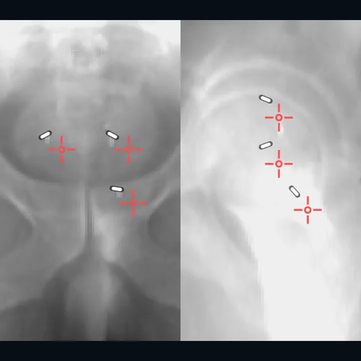
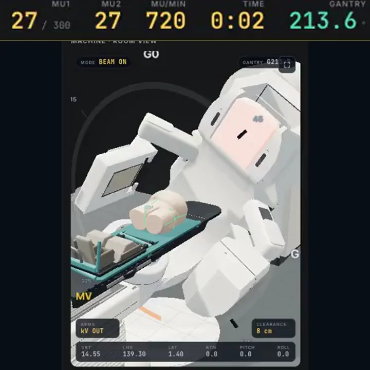
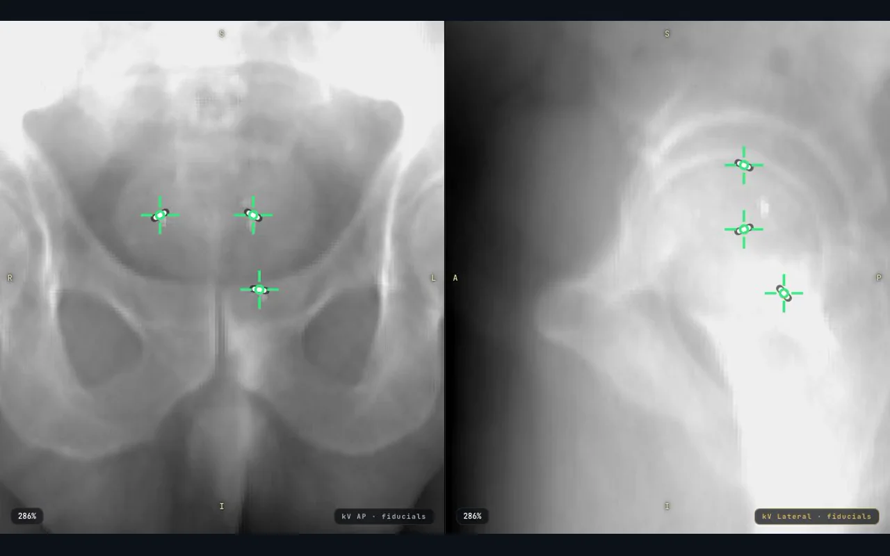
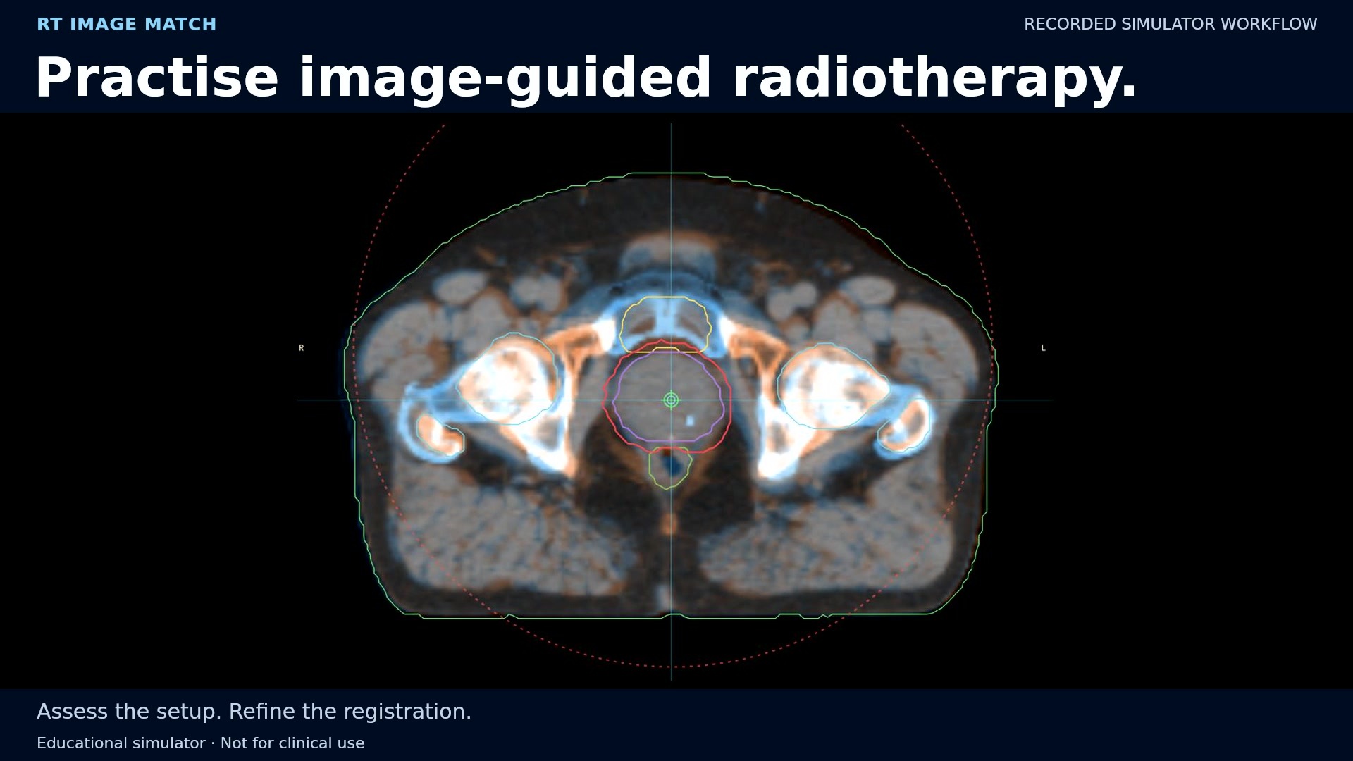
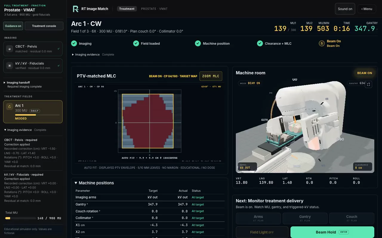
The scroll animation retains the original September 9 CBCT alignment and paired fiducial-verification frames. The Deliver scene is a new October 6 recording of the actual Prostate VMAT Full Treatment simulator, with the current training LINAC, changing MLC aperture, gantry rotation and increasing simulated monitor units. It was captured in the existing local fictional-case fixture. Image-acquisition waits and guide placement are condensed; actual imaging completion, held couch correction, field preparation, collision checks and Beam On remain active. The earlier matching footage and new delivery footage are separate recording sessions. The example ends in a brief triggered-imaging hold, not field or fraction completion.
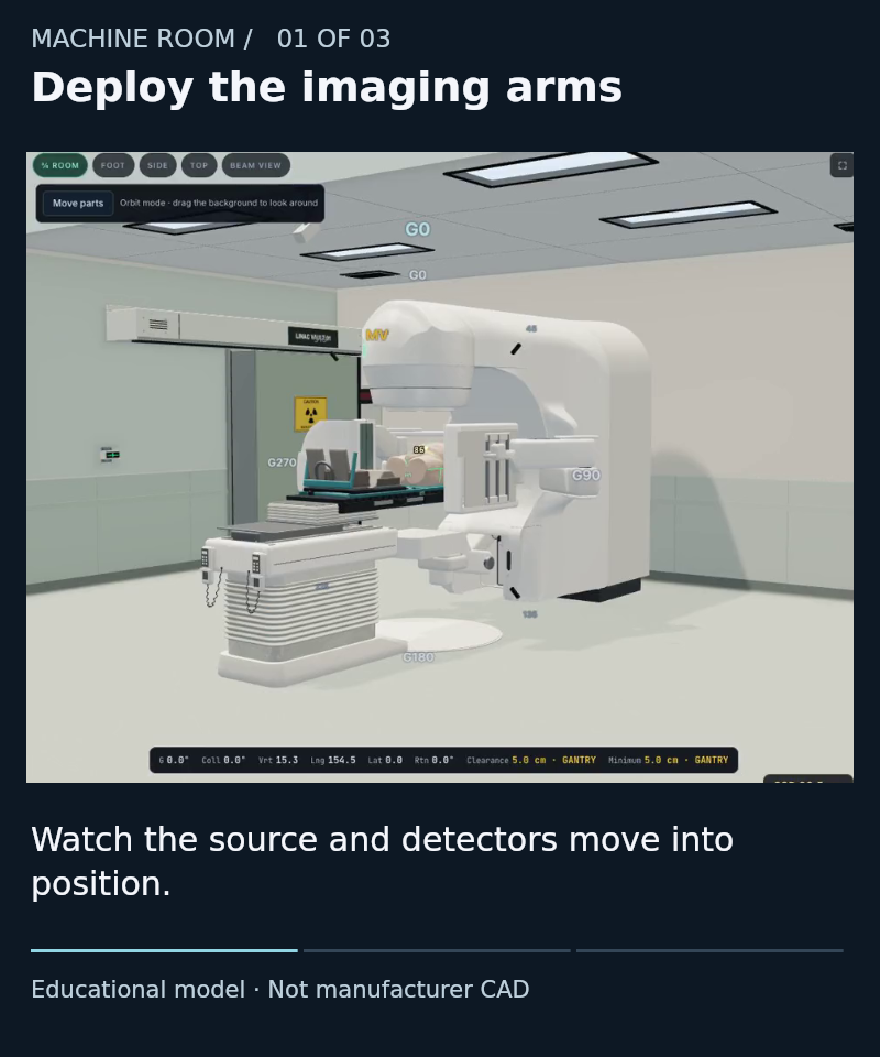
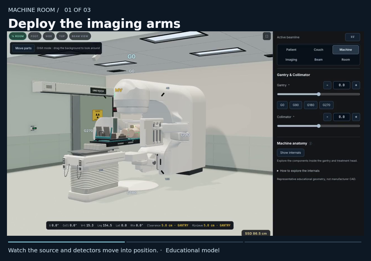
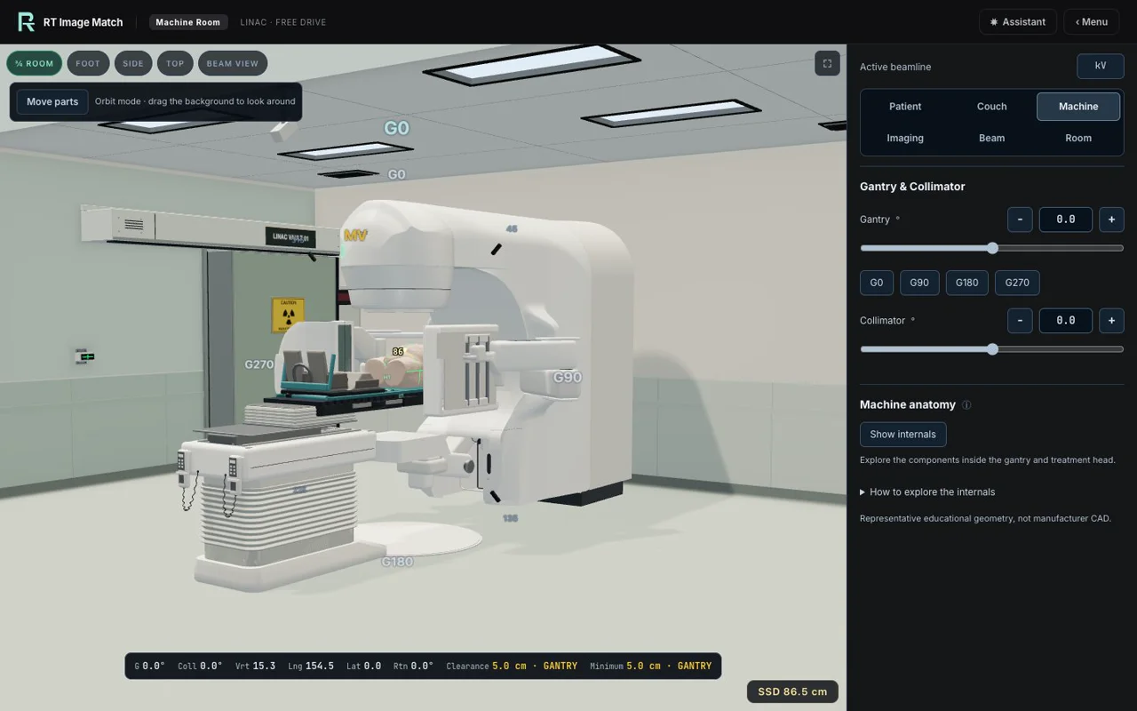
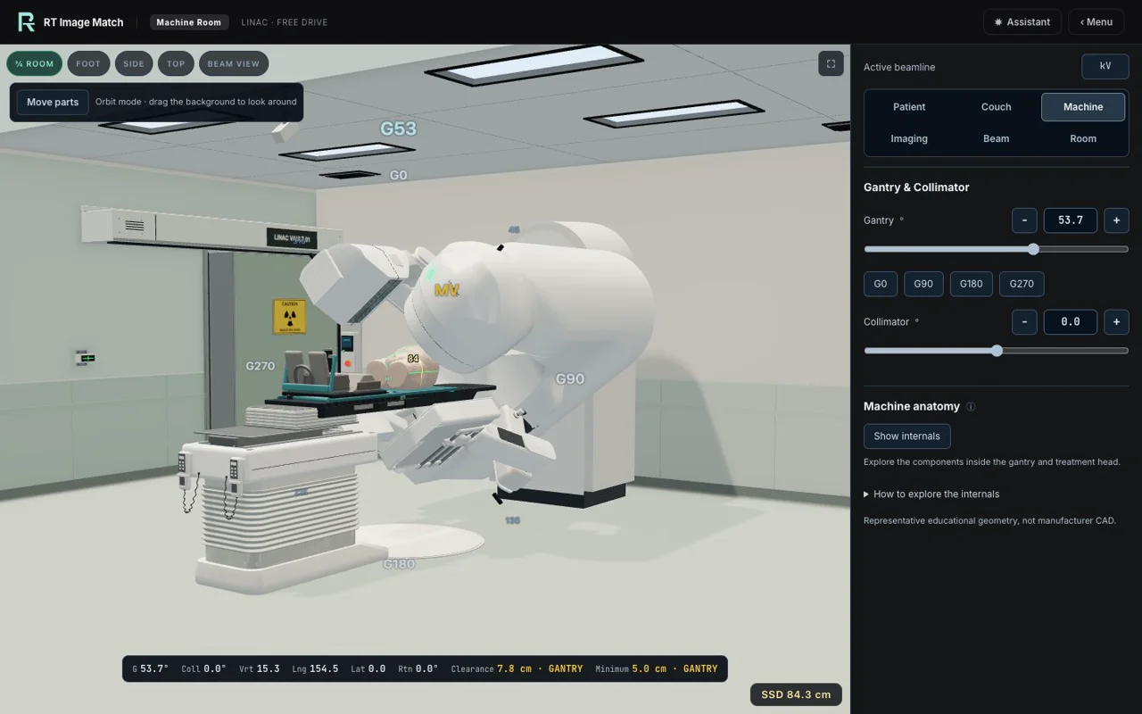
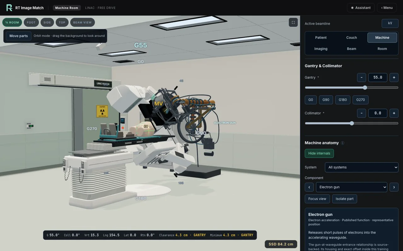
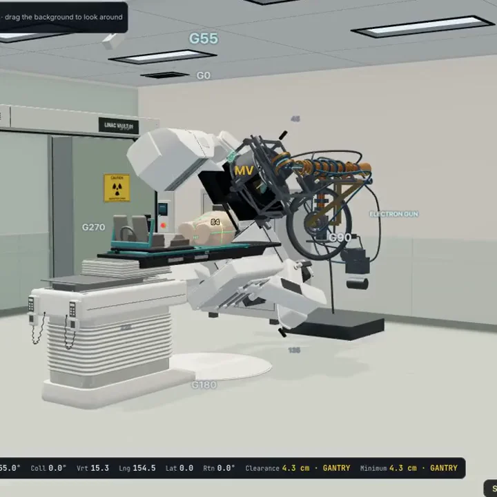
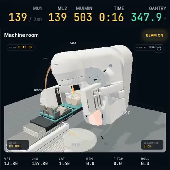
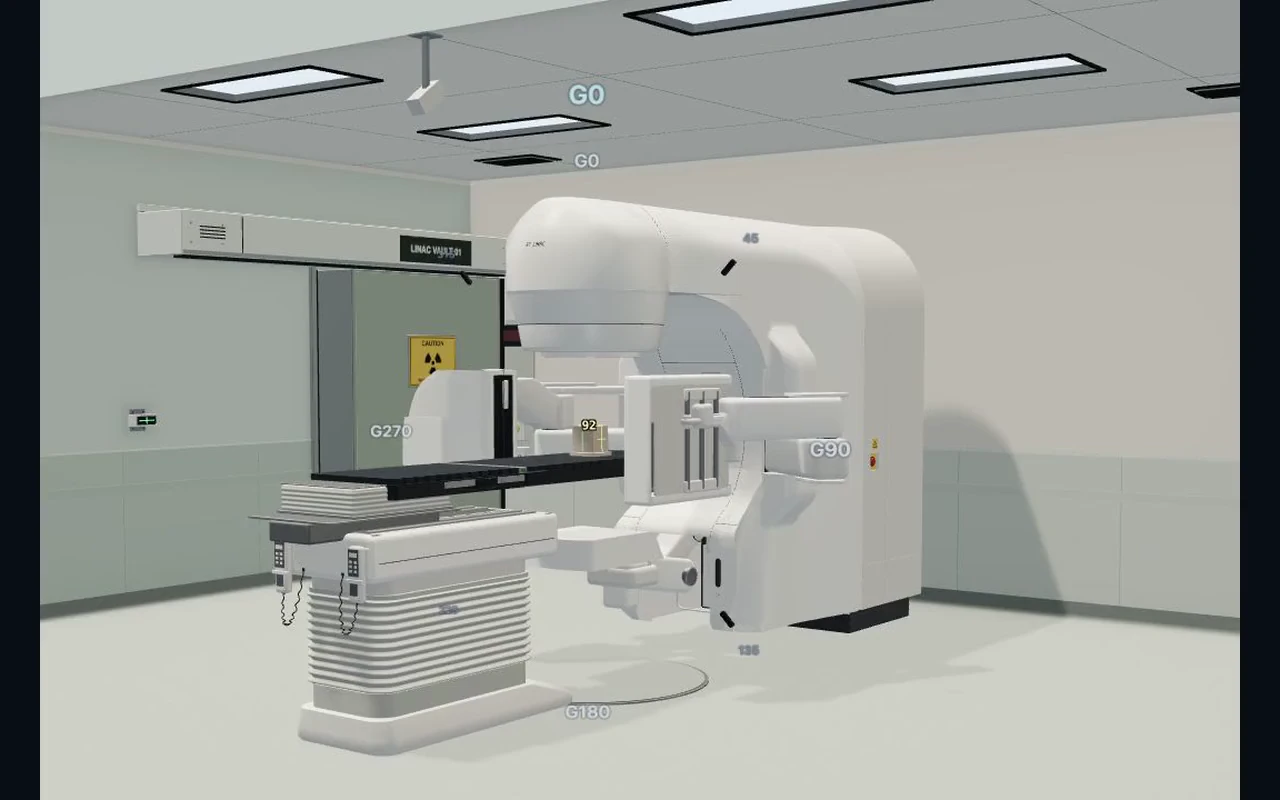
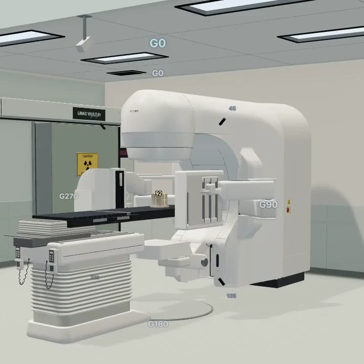
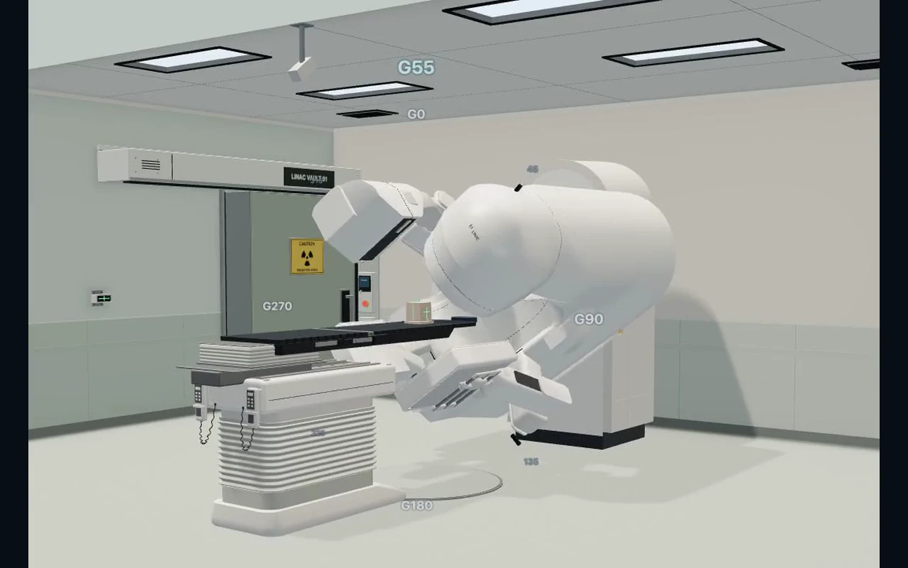
October 6 Machine Room captures show actual imaging-arm deployment and gantry rotation, then a whole-gantry internal reveal at 55°. The casing is hidden through the native controls while the accelerator and beam path remain in the context of the complete machine. The new fullscreen crop differs slightly from the preceding rotation view. The QA phantom and equipment are representative teaching geometry, not manufacturer CAD.
Timing is edited, not real time. Seven seconds of captured Full Treatment delivery are retimed to ten seconds, omitting the long imaging-hold tail. Native whole-gantry screenshots are sparse and condensed to four seconds. Silent 24-frame-per-second files select or repeat recorded frames; no optical interpolation or generated intermediate motion is added. Phone versions reframe the original machine view and treatment readouts. No source head or facial imaging, private learner records or account details appear. The pelvic source is Prostate-Anatomical-Edge-Cases (TCIA), licensed CC BY 4.0; changes were made. Values and setup are fictional. Educational use only, not a clinical protocol.
Current scroll recordings and endpoint images
- treatment-story-current-20261006.mp4
- treatment-story-current-20261006-mobile.mp4
- machine-story-current-20261006.mp4
- machine-story-current-20261006-mobile.mp4
- treatment-delivery-current-20261006.webp
- treatment-delivery-current-20261006-mobile.webp
- machine-arms-current-20261006.webp
- machine-arms-current-20261006-mobile.webp
- machine-gantry-current-20261006.webp
- machine-gantry-current-20261006-mobile.webp
- machine-internals-current-20261006.webp
- machine-internals-current-20261006-mobile.webp
Case-library screenshots
The landing-page library lists all 39 current case and workflow entries: 13 planar, 16 CBCT/MR, and ten Full Treatment workflows. Its 38 image-backed entries use 37 complete reviewed trainer stills and two focused phone crops, captured in an isolated local fixture with fictional setup values and no real learner records. Thumbnails reuse the same complete images. The Partial Breast DIBH entry explicitly reuses its existing partial-breast CBCT reference anatomy; it does not show DIBH delivery. The Whole Brain entry is listed without a preview image.
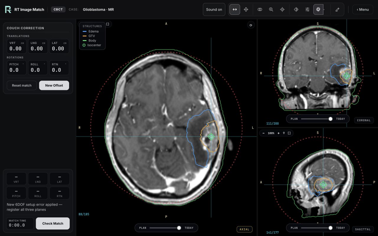
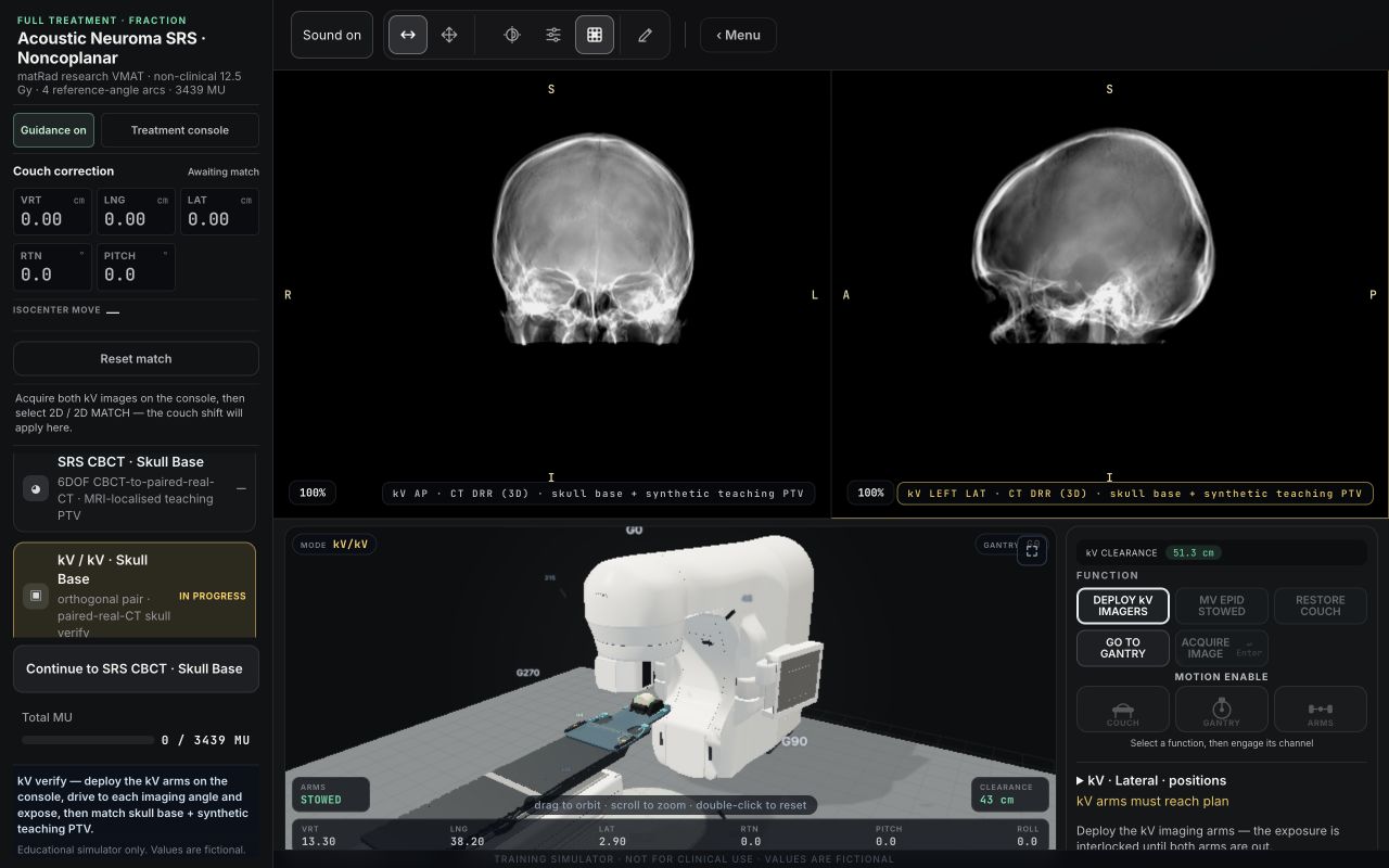
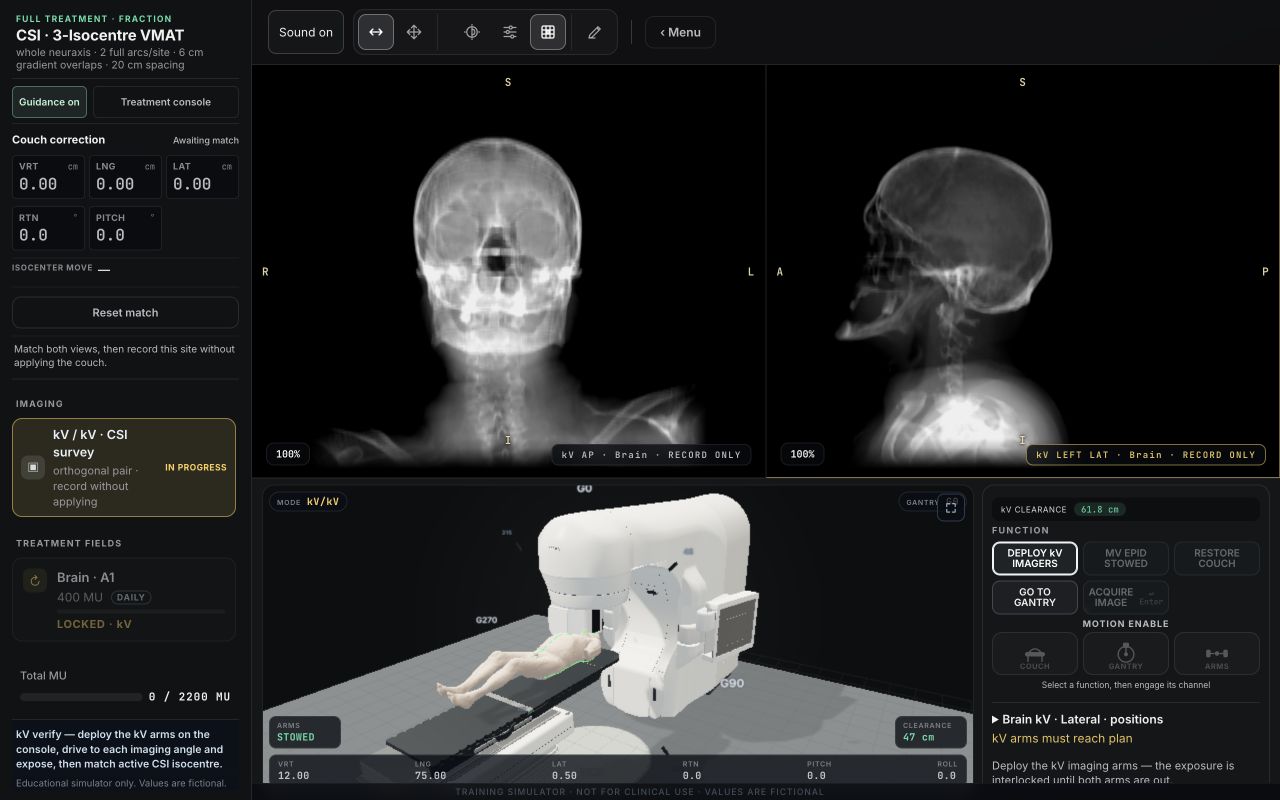
The original 30 images retain their recorded provenance and WebP encoding. Six of those show Full Treatment matching with the linac in the same frame: CBCT for Prostate, Lung and Liver; paired kV for Instrumented Spine and Breast; and the synthetic five-BB Machine QA phantom. Acquisition timing was condensed for those stills. On 7 October 2026, seven head-case previews were added at the site owner's request as unmodified native 1280 × 800 JPEG screenshots. The two native Full Treatment views show reference DRRs and the linac during kV acquisition preparation: CSI's Brain station and Acoustic Neuroma's optional skull verification. They do not show acquired-image approval or completed delivery. The Acoustic Neuroma still is now labelled as an earlier skull-verification workflow; the current Cranial SRS case uses PROTEAS P28 and is not pictured in that capture.
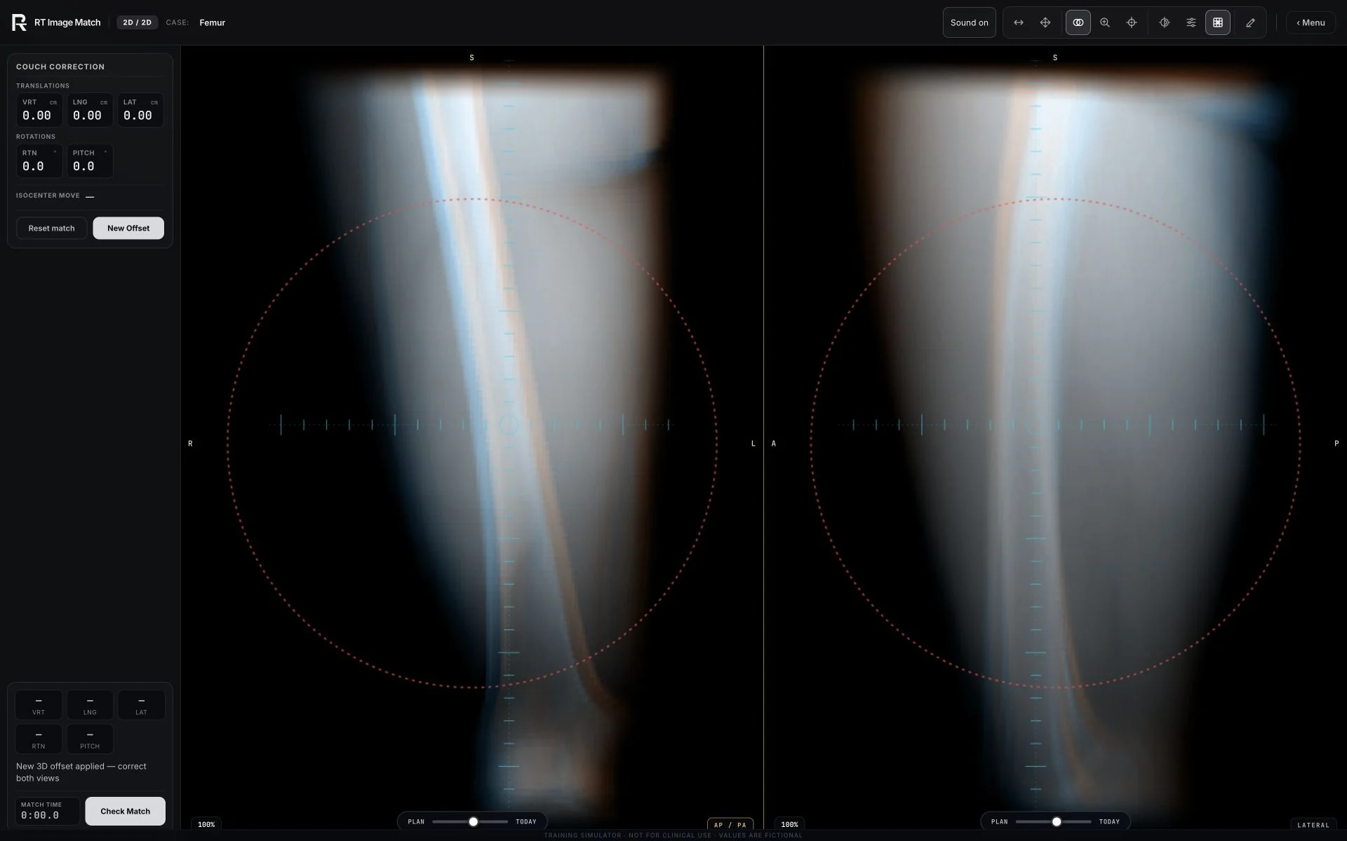
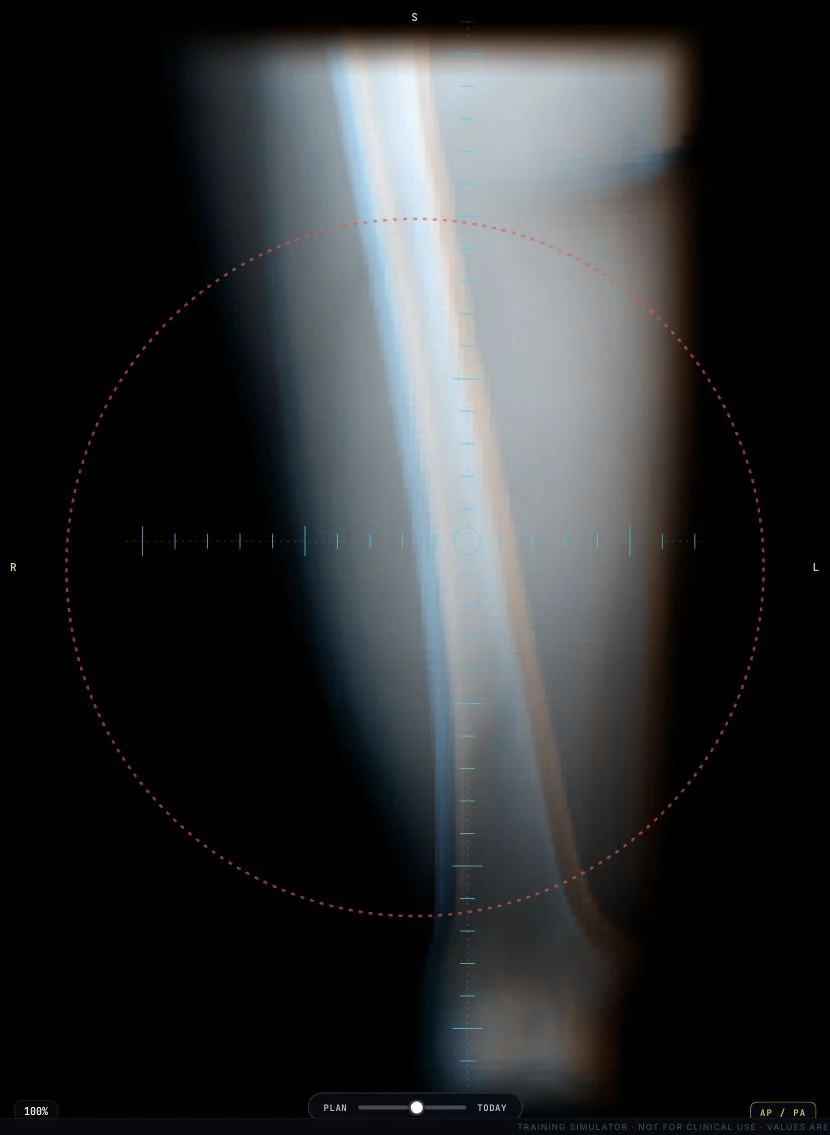
On 15 October 2026, Femur and Mediastinal Lymph Nodes were recaptured from the actual matching workspace at 1920 × 1200. The Femur phone view shows its AP pane; the Mediastinal phone view shows its axial pane. These native screenshot crops retain the real image overlays and blend control. Enlarge displays the complete workspace. Only WebP compression was applied; no anatomy was generated, retouched or upscaled. The views show the thigh and thorax, respectively, with fictional setup errors and no submitted grade. Their sources remain Soft-tissue-Sarcoma and Mediastinal-Lymph-Node-SEG below. The share image uses the same reviewed axial crop with the site's RT mark and typography.
Earlier gallery captures
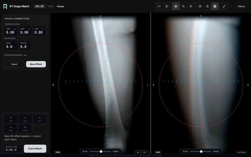
The earlier Femur and Mediastinal Lymph Nodes views remain available for comparison with their original capture records. They have not been relabelled as new captures.
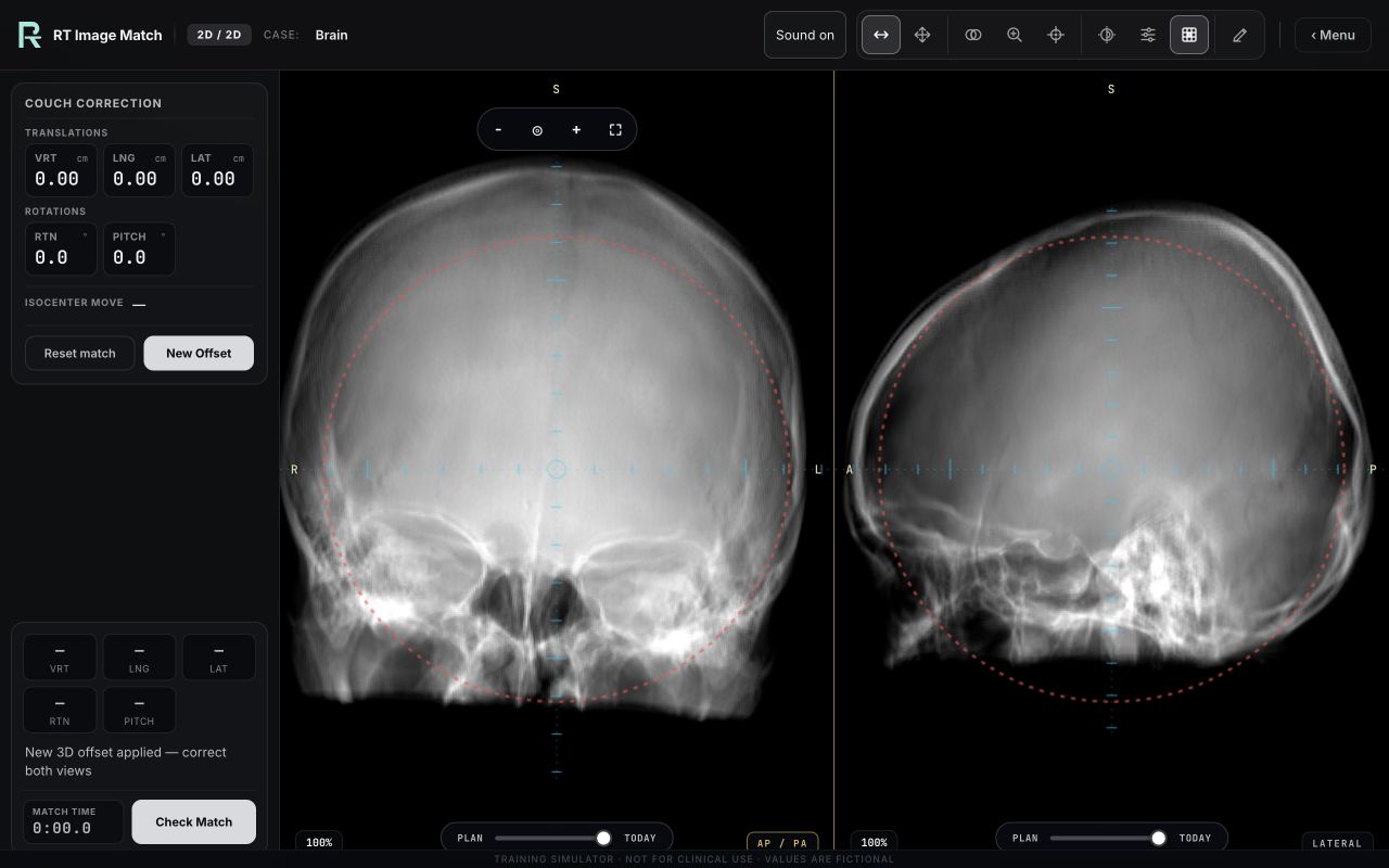
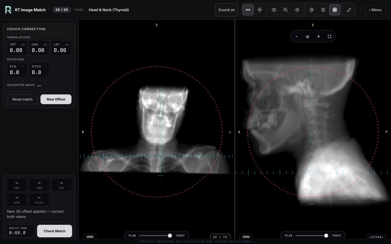
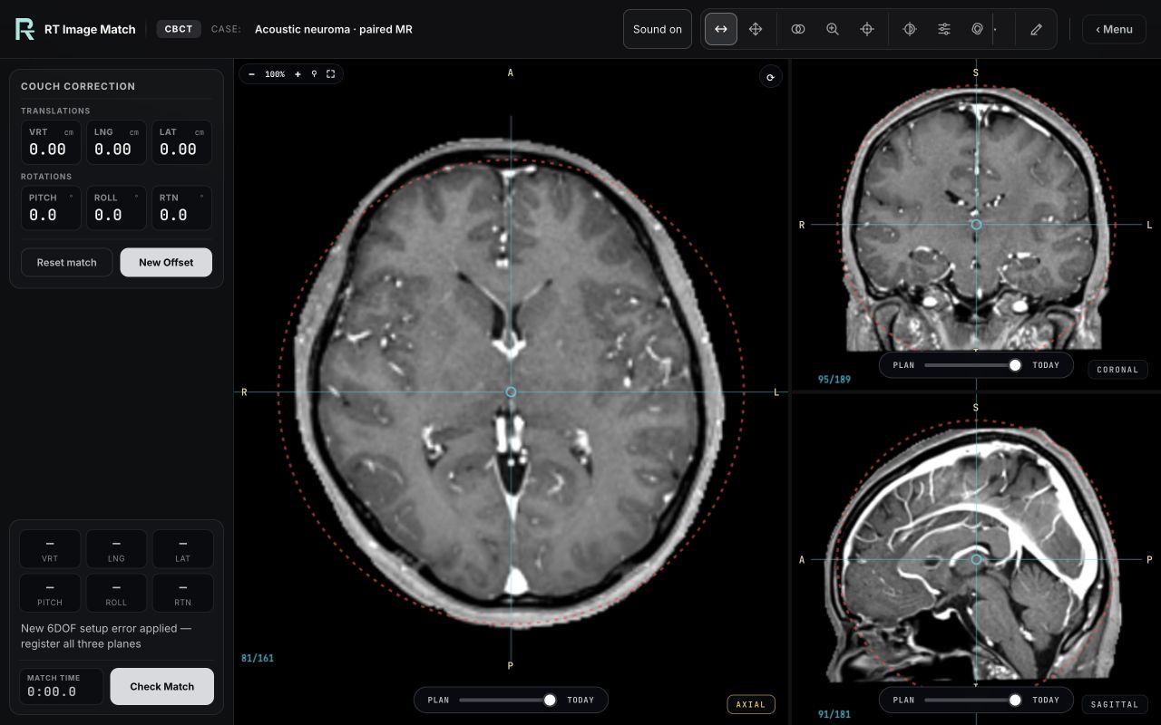
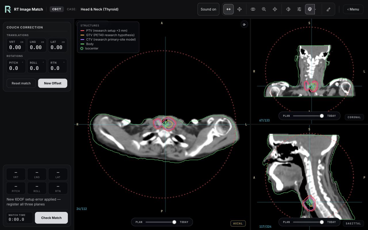
These public previews include research-derived head anatomy. No direct identifiers or real learner records are shown. The underlying case datasets remain protected. Publication does not establish source-specific consent, ethics approval, de-identification, or facial-data reuse clearance; the source limitations and TCIA restrictions below still apply. No montage or AI-generated patient imagery was added to the captures. Each image is mapped to its sources and exact hash in the image-rights register. These are non-clinical educational previews, not completed treatments or clinical QA measurements.
Restored head-case stills: Brain, Head & Neck (Thyroid), Acoustic neuroma · paired MR, Glioblastoma · MR, Head & Neck (Thyroid), Acoustic Neuroma SRS · Noncoplanar, CSI · 3-Isocentre kV.
Anatomy locator and viewing planes
The body locator, selective bone landmarks and viewing-plane guide are original schematic illustrations created for this site. The face-free body was authored in Blender from deterministic cross-sections and shallow shoulder, pelvic, knee, hand and foot landmarks. The femurs, lower legs, pelvis and segmented spine use separately authored simplified geometry. Their generator, geometry counts and hash are recorded in the project. Selected areas become translucent to explain location; these are not patient segmentations, organ contours or treatment targets. The guide contains no source-patient surface, case volume, imported model or manufacturer geometry. Recorded case screenshots remain separate from the illustrations.
Imaging datasets
The nine active Full Treatment patient cases use fictional courses: prostate 60 Gy/20 fractions, lung 60 Gy/30, cranial SRS 20 Gy/1, liver metastasis 50 Gy/5, instrumented spine 20 Gy/5, whole breast 40.05 Gy/15, partial breast 30 Gy/5, whole brain 30 Gy/10 and adult craniospinal phase 36 Gy/20. Six include independently calculated educational plans, contours, organs at risk and native-grid cumulative dose-volume histograms: lung, cranial SRS, instrumented spine, whole breast, partial breast and whole brain. Their delivered fields come from those plans: lung and partial-breast dynamic IMRT, SRS arcs and couch angles, the spine and whole-brain static fields and the whole-breast field-in-field segments. Prostate, liver and craniospinal deliver illustrative VMAT arcs, and their plan review states that dose and DVH are unavailable. Original setup imaging is retained. Display resampling does not alter native DVHs. Automatic and authored contours, respiratory-motion gaps, unassessed CSI coverage and remaining planning goals are explicitly identified. These are not source patient prescriptions, clinical dose calculations or approved treatment plans.
Trainer cases are built from the following collections, accessed through The Cancer Imaging Archive (TCIA) / the NCI Imaging Data Commons unless noted. This inventory includes every DOI and CC BY source embedded in still-distributed case-data files or protected DRRs and every source visible in a public trainer capture, including assets whose start-screen card has been retired or that are now used only in challenges.
- Prostate Anatomical Edge Cases Prostate fiducial CBCT case and 2D/2D prostate fiducial-match case (planning CT with the patient's real implanted gold markers); and the 2D/2D Hip Prosthesis / Pelvis case from O-MAR planning CT patient Prostate-AEC-111 (real unilateral total-hip arthroplasty). AP and patient-left lateral teaching DRRs were rendered from the CT; support hardware was removed from the projection volume. The Full Treatment case delivers three illustrative full VMAT arcs for a fictional interim prescription of 60 Gy in 20 fractions; its plan review states that dose and DVH are unavailable. Original setup imaging is retained. This is not the source patient's treatment plan or a clinically approved plan; remaining planning and contour limitations are displayed. Changes were made. The Cancer Imaging Archive — doi:10.7937/qstf-st65 · CC BY 4.0
- Pediatric-CT-SEG 2D/2D and CBCT Pediatric Abdomen cases from exam Pediatric-CT-SEG-E1FF3C7E. The near-isotropic CT was converted into AP and patient-left lateral educational DRRs and an abdomen/pelvis isotropic CBCT atlas. The CBCT case preserves selected expert Skin, liver, kidney, stomach, bladder, spinal-canal and bone contours as a non-cancer organ-registration exercise; no target volume was added. Changes were made. The Cancer Imaging Archive — doi:10.7937/TCIA.X0H0-1706 · CC BY 4.0
- LIDC-IDRI + DICOM-LIDC-IDRI-Nodules Breast CBCT case (synthetic lumpectomy plan, cavity, and clips added), also reused unchanged for the Partial Breast L · DIBH IMRT Full Treatment workflow (LIDC-IDRI-0558; a static teaching breath-hold reference, not paired DIBH imaging; the earlier geometric SlicerRT lesson is superseded in Full Treatment by an independently calculated five-field sliding-window dynamic IMRT 6 MV plan with fictional 30 Gy in five nonconsecutive fractions and native DVHs; its same-CT automatic organs, heart and breast tissue come from TotalSegmentator 2.16.0’s open
totalandbreaststasks and are not clinician-reviewed); the 2D/2D Breast L mono-isocentric supraclavicular + medial-tangent case (reference field shapes baked into the adapted images); and a legacy Lung SBRT CBCT atlas that remains in the protected case-data inventory. That lung atlas uses released research CT subject LIDC-IDRI-0136 and a strict 2-of-3 consensus of the exact three separately published DICOM Nodule 7 reader annotations. This site does not independently assert a source-specific de-identification, consent, ethics, or re-identification status for that CT. It preserves the visible source lesion and CT-derived body/lung structures and adds a synthetic 5 mm teaching PTV. It is not the current 4D-Lung case, but its assets are still distributed and are therefore credited here. Changes were made. Source CT — The Cancer Imaging Archive — doi:10.7937/K9/TCIA.2015.LO9QL9SX · CC BY 3.0 Nodule annotations — The Cancer Imaging Archive — doi:10.7937/TCIA.2018.H7UMFURQ · CC BY 3.0 - CMB-BRCA (Cancer Moonshot Biobank — Invasive Breast Carcinoma) Protected planning DRRs for the Breast Supine tangent exercise were rendered from patient MSB-02054. The CT was cropped and converted into educational projection images; setup errors and field interaction are simulated, and changes were made. The Cancer Imaging Archive — doi:10.7937/DX22-8J71 · CC BY 4.0
- 4D-Lung Standalone Lung matching and Full Treatment conventional NSCLC cases (patient 100_HM10395: one 50% / end-exhale planning 4DCT is used for both Plan and Today, with a fictional rigid 6DOF setup error applied to Today; the physician planning RTSTRUCT tumour/lung/cord/heart contours were rasterized, and a synthetic PTV of GTV + 5 mm was added); the CT Simulation Chest / lung exercise from arms-up patient 110_HM10395 (all ten matched 0–90% gated source CT phases across the finite pelvis/abdomen-through-lower-neck acquisition, downsampled to one common teaching grid; BODY and lung masks are automated derivatives, not physician contours; no target, plan, dose, or synthetic anatomy continuation was added); and the Breast L · Opposed Tangents 3D Conformal Full Treatment case from the arms-up 0% planning phase of patient 106_HM10395. For the breast case, the source RTSTRUCT lungs, heart, cord, and esophagus were used for geometric review; TotalSegmentator-derived breast anatomy was used to author an explicitly synthetic whole-breast teaching target; Plastimatch rendered setup and portal projections; and the patient-specific equal-and-opposite collimator rotations plus exact two-field jaw/MLC geometry were instantiated and read back in SlicerRT. No source breast treatment plan or dose was used. Changes were made. The Cancer Imaging Archive — doi:10.7937/K9/TCIA.2016.ELN8YGLE · CC BY 3.0
- NSCLC Radiogenomics 2D/2D Thorax case (AP + patient-left lateral planning DRRs generated from a chest CT). The Cancer Imaging Archive — doi:10.7937/K9/TCIA.2017.7HS46ERV · CC BY 3.0
- TCGA-PRAD (The Cancer Genome Atlas — Prostate Adenocarcinoma) 2D/2D Pelvis case (bony DRRs ray-summed from a diagnostic chest/abdomen/pelvis CT of patient TCGA-VP-A878). This collection also supplied MV-style pelvis and lumbar portal renders that remain in the case-data file but are not currently exposed as trainer cases; they are credited here because they are still distributed. The separate 2D/2D Lumbar Spine case is not derived from this collection — see Spine-Mets-CT-SEG below. Changes were made. The Cancer Imaging Archive — doi:10.7937/K9/TCIA.2016.YXOGLM4Y · CC BY 3.0
- Soft Tissue Sarcoma Soft-tissue sarcoma CBCT case (real tumour contour), 2D/2D femur case, and 2D/2D Shoulder / Scapula case from left-shoulder patient STS_008. The public 2D/2D landing poster is a deterministic capture of patient STS_004's cropped single-thigh Femur case; it contains no head or facial anatomy. The shoulder planning references were centred on the real GTV and converted into educational DRRs. The 2D/2D Knee / Tibia case uses whole-leg patient STS_047: the contralateral leg was removed at the measured inter-leg gap and the field was centred on the femoral condyles, with the released GTV_Mass ROI used only to anchor that search. Changes were made. The Cancer Imaging Archive — doi:10.7937/K9/TCIA.2015.7GO2GSKS · CC BY 3.0
- Spine-Mets-CT-SEG Spine Metastasis T4 2D/2D, CBCT and Full Treatment surfaces from patient 13683 use the same 0.5 mm radiotherapy-simulation CT and released T4 vertebral DICOM-SEG. Their start-screen cards have been retired, so those surfaces are reachable only through the rotating challenge; the assets remain in the case data and are credited here because they are still distributed. The collection classification identifies an osteolytic T4 level extending into the pedicle and pars; no lesion contour or patient treatment plan is released. Same-patient AP/lateral and beam-angle planning references were generated with Plastimatch. A 3 mm BODY-clipped expansion of the source T4 structure, carved outside a 2 mm cord-avoidance envelope, was created and read back in 3D Slicer. Three posterior static fields, jaws and a 120-leaf geometric MLC aperture with a separate 5 mm allowance were instantiated and read back in SlicerRT. That PTV, all field geometry, MU and delivery are explicitly synthetic educational simulation; no optimization, prescription, dose calculation or deliverable patient plan is represented. The separate Instrumented Spine 2D/2D and simulated first-day workflow use patient 11471 with a real three-level thoracic pedicle-screw and rod construct; their T6–T8-derived target and SlicerRT-read-back AP/PA plan are likewise synthetic. The separate Lumbar Spine 2D/2D case uses patient 10456, a 0.5 mm skin-to-skin radiotherapy-simulation CT whose released vertebral DICOM-SEG spans T8–L5; the L3 segment supplies the isocentre for AP/lateral Plastimatch references and no target volume, margin or plan is derived from it. Changes were made. The Cancer Imaging Archive — doi:10.7937/KH36-DS04 · CC BY 4.0
- TCGA-THCA (The Cancer Genome Atlas — Thyroid Cancer) Head & neck 2D/2D and CBCT cases from patient TCGA-DE-A4MA. DRRs were ray-summed from the head-and-neck CT. For the research-only CBCT teaching case, TotalSegmentator 2.16.0 generated a thyroid-gland mask and both lobes were reviewed in 3D Slicer 5.10. The same-study PET40 focus is used only as a gross-disease hypothesis (GTV, 6.0 cc). A simplified primary-site CTV (27.0 cc) combines the reviewed thyroid with a Slicer +5 mm local GTV expansion, then uses a Slicer +7/−7 mm morphological close to restore a connected isthmus region. One connected PTV (44.8 cc) encompasses both lobes with a Slicer +3 mm setup expansion. No nodal CTV is invented. These are literature-informed research teaching volumes—not released physician contours, a prescription, dose input, optimisation or deliverable plan. Changes were made. The Cancer Imaging Archive — doi:10.7937/K9/TCIA.2016.9ZFRVF1B · CC BY 3.0
- CPTAC-UCEC + CPTAC-UCEC-Tumor-Annotations Gynae / uterus CBCT case from contrast pelvic CT patient C3N-00872. The separately published exact RTSTRUCT ROI
UTERUS - 1is a RECIST-style radiology lesion/tumour annotation labelled by organ; it is preserved as the source registration target but is not a whole-organ uterus contour or RT GTV. Bladder and rectum are derived teaching contours. The trainer presents a rigid 6DOF registration rather than synthetic differential target motion. Two nested BODY-clipped target volumes were made and read back with the 3D Slicer 5.10 Segment Editor Margin effect as requested 5 mm + 5 mm steps. They are explicitly synthetic CTV/PTV teaching geometry, not clinician contours, dose inputs, optimization, or a deliverable plan. Changes were made. Source CT — The Cancer Imaging Archive — doi:10.7937/K9/TCIA.2018.3R3JUISW · CC BY 4.0 Lesion annotation — The Cancer Imaging Archive — doi:10.7937/89M3-KQ43 · CC BY 4.0 - CC-Tumor-Heterogeneity Cervical Cancer CBCT-mode case from patient CCTH-A03's pre-treatment Timepoint1 acquisition. A source cervical-tumour annotation drawn on the collection's T2-weighted MRI was mapped to its paired pelvic CT through the released rigid DICOM Spatial Registration object. The CT was body-masked, cropped and resampled for the trainer; the moving cone-beam view is simulated from that CT rather than an independent daily scan. Two nested BODY-clipped geometric teaching contours were generated from the unchanged source annotation with the 3D Slicer 5.10 Segment Editor Margin effect as requested 5 mm + 5 mm steps. They are explicitly synthetic and are not represented as GTV, CTV, ITV, PTV, clinician-delineated treatment margins, dose inputs, optimization, or a plan. Changes were made. The Cancer Imaging Archive — doi:10.7937/ERZ5-QZ59 · CC BY 4.0
- Mediastinal-Lymph-Node-SEG TotalSegmentator 2.16.0 added automated trachea, esophagus, aorta and combined lung contours from the original CT. Tiny disconnected lung islands and contours on the atlas's blank end slices were omitted; all original nodes and teaching margins were preserved. Mediastinal Lymph Nodes CBCT-mode case from fully annotated contrast CT patient case_0412. All four components of the source manual lymph-node DICOM-SEG were retained while the CT was body-masked, cropped and resampled; the moving cone-beam view is simulated from that CT rather than an independent daily scan. The trainer presents the nodes and thoracic anatomy as one rigid 6DOF registration. Two nested BODY-clipped geometric teaching contours were generated from the unchanged diagnostic annotation with the 3D Slicer 5.10 Segment Editor Margin effect as requested 5 mm + 5 mm steps. The expansions are explicitly synthetic and are not represented as GTV, CTV, ITV, PTV, clinician-delineated treatment margins, dose inputs, optimization, or a plan. Changes were made. The Cancer Imaging Archive — doi:10.7937/QVAZ-JA09 · CC BY 4.0
- CPTAC-SAR (Clinical Proteomic Tumor Analysis Consortium — Sarcoma) CSI Full Treatment and Machine Room patient from patient C3N-00875's continuous whole-body CT. Planning AP and patient-left lateral teaching DRRs were generated at Brain, Superior Spine, and Inferior Spine stations exactly 200 mm apart; the patient support was removed from the projection volume and the images were cropped, ray-summed, contrast-windowed, and resampled. Machine Room brain, lungs, heart, liver, kidneys, bladder, and spinal-cord masks were generated on this same CT with TotalSegmentator 2.15.0 and resampled to the room atlas. These structures remain available in the explicit 3D cutaway beneath the feature-suppressed outer head surface. The available structures are automated teaching segmentations, not physician contours or treatment-planning structures. All setup errors, couch parameters, 0.5/0.7 cm scenario margins, treatment sequence, and delivery are synthetic educational simulation only; no source patient plan, target, dose, or clinical protocol is represented. Changes were made. The Cancer Imaging Archive — doi:10.7937/TCIA.2019.9BT23R95 · CC BY 4.0
- CPTAC-CM (Clinical Proteomic Tumor Analysis Consortium — Cutaneous Melanoma) Female Whole-Body Machine Room patient from patient C3N-02633's continuous head-first CT. The scanner cradle was removed with a deterministic source-CT BODY mask, the complete acquired head-to-feet extent was retained, and the room atlas was resampled to 4 mm. Brain, lungs, heart, liver, kidneys, bladder, spinal cord, and breast tissue were generated on this same CT with TotalSegmentator 2.15.0 and resampled to the exact room grid. These structures remain available in the explicit 3D cutaway beneath the feature-suppressed outer head surface; the available structures are automated teaching segmentations, not physician contours or treatment-planning structures. The TotalSegmentator 2.15.0 models used here do not provide this case's ureters, urethra, uterus/cervix, ovaries, fallopian tubes, or vagina; those layers are clearly labelled, non-patient-specific educational schematics anchored to the same-scan kidneys and bladder. The patient is provided only for linac-room setup, movement, collision, laser, ODI, imaging, accessory, tattoo, and cutaway practice; no plan, target, prescription, dose, treatment sequence, or clinical protocol is represented. Data used in this project were generated by the National Cancer Institute Clinical Proteomic Tumor Analysis Consortium (CPTAC). Changes were made. The Cancer Imaging Archive — doi:10.7937/K9/TCIA.2018.ODU24GZE · CC BY 4.0
- Pancreatic-CT-CBCT-SEG (Hong J. et al.) Pancreas CBCT case (breath-hold planning CT with gastrointestinal organ contours). The Cancer Imaging Archive — doi:10.7937/TCIA.ESHQ-4D90 · CC BY 4.0
- Colorectal-Liver-Metastases + BAMF-AIMI Annotations Liver SBRT CBCT and teaching-workflow case from patient CRLM-CT-1100. The collection contrast CT and DICOM-SEG supply the source anatomy plus hepatic and portal vessel contours. The exact BAMF-AIMI AI-generated DICOM-SEG labelled “AIMI liver-tumor radiologist 7 corrected segmentation” supplies the corrected liver and Mass contours used by the trainer. The images were cropped, resampled, projected and display-windowed, and changes were made. The displayed CTV/PTV layers are synthetic teaching overlays, not source contours, clinical margins, dose, or a deliverable plan. Source CT, hepatic/portal annotations — The Cancer Imaging Archive — doi:10.7937/QXK2-QG03 · CC BY 4.0 Corrected liver/Mass annotation — Zenodo — doi:10.5281/zenodo.8345959 · CC BY 4.0
- UPenn-GBM + BAMF-AIMI Annotations Glioblastoma MR case from UPENN-GBM patient UPENN-GBM-00019. The post-contrast T1 source MR is paired with the exact BAMF-AIMI AI-generated DICOM-SEG labelled “AIMI Brain MRI radiologist 3 corrected segmentation.” The enhancing-lesion and necrosis segments were combined into the displayed tumour target and the edema segment was retained for context; the external head contour was derived from the MR. The MR was cropped and display-normalised, the corrected SEG was reoriented and resampled to the teaching atlas, and changes were made. The underlying MR data remains protected; one individually reviewed head-case still is used in the public case library. Source MR — The Cancer Imaging Archive — doi:10.7937/TCIA.709X-DN49 · CC BY 4.0 Tumour annotation — Zenodo — doi:10.5281/zenodo.8345959 · CC BY 4.0
- Adrenal-ACC-Ki67-Seg Adrenal CBCT case from a contrast abdominal CT with a radiologist-refined adrenal-mass DICOM-SEG. TotalSegmentator 2.16.0 added automated left/right kidney, spleen and stomach anatomy from the exact CT used by the trainer. Tiny disconnected prediction islands were removed; the existing CT and source mass were unchanged. The source mass is preserved and registered with the surrounding abdominal anatomy as one rigid 6DOF volume. Two nested BODY-clipped geometric teaching contours were generated from the unchanged source annotation with the 3D Slicer 5.10 Segment Editor Margin effect as requested 5 mm + 5 mm steps. The expansions are explicitly synthetic and are not represented as GTV, CTV, ITV, PTV, clinician-delineated treatment margins, dose inputs, optimization, or a plan. Changes were made. The Cancer Imaging Archive — doi:10.7937/1FPG-VM46 · CC BY 4.0
- CPTAC-CCRCC + CPTAC-CCRCC-Tumor-Annotations Renal Cell Carcinoma CBCT case from patient C3N-03018. Left and right kidney anatomy was added with TotalSegmentator 2.16.0 on the original acquisition-1 CT and resampled into the unchanged teaching grid; these are automated teaching contours, not physician organ contours. The contrast CT was cropped and resampled into an isotropic teaching atlas; the radiologist-reviewed right-kidney tumour annotation is preserved as a source lesion outline. It is not represented as a clinical GTV, CTV or PTV, and no treatment margin was inferred. Changes were made. Source CT — The Cancer Imaging Archive — doi:10.7937/K9/TCIA.2018.OBLAMN27 · CC BY 4.0 Tumour annotation — The Cancer Imaging Archive — doi:10.7937/SKQ4-QX48 · CC BY 4.0
- EAY131 + EAY131-Tumor-Annotations v2 Esophageal Cancer CBCT case from patient EAY131-8537265. The thoracic CT was gap-filled from its released non-uniform slice positions, cropped and resampled into an isotropic teaching atlas; the expert esophageal-primary annotation is preserved as a source lesion outline. The 3D Slicer 5.10 Segment Editor Margin effect added requested +5 mm and +10 mm-total BODY-clipped synthetic teaching contours with SlicerRT verified. The outer contour is shown in the trainer’s synthetic PTV row, but neither expansion is a clinician-delineated GTV, CTV, ITV or PTV, clinical margin, dose input, optimization result, or treatment plan. Changes were made. Source CT — The Cancer Imaging Archive — doi:10.7937/C5KE-YX42 · CC BY 4.0 Tumour annotation — The Cancer Imaging Archive — doi:10.7937/Q9RN-M510 · CC BY 4.0
- PROTEAS — Brain Metastases Cranial SRS Full Treatment uses P28 planning CT and source RTSTRUCT BODY/PTV on an exact shared 2 mm grid. The simulated daily CBCT and optional skull kV images are derived from that same planning CT; they are not separate patient acquisitions. Browser window quantization, PNG packing, complete synthetic head exterior, fictional daily setup and an independently generated four-arc research plan were added. The current fictional course is 20 Gy in one fraction, independently calculated on a 1 mm dose grid with native DVHs and a separate 2 mm display. The prior 12.5 Gy research baseline is retained separately; neither prescription is inferred from the source PTV20Gy name. Whole Brain 3D Conformal uses P30 CT and source brain, brainstem, cerebellum, eye, lens and BODY contours, a derived whole-brain target with 3 mm PTV, linked kV and divergent MV ports, and a separate fictional 30 Gy / 10 fraction opposed-lateral matRad plan with uncommissioned model MU. No MRI registration or clinical treatment validation is claimed. Zenodo — doi:10.5281/zenodo.23171182 · CC BY 4.0
- A Paired Head CT-MRI Dataset for Cross-Modality Image Synthesis 2D/2D Brain and standalone Acoustic neuroma paired-MR practice use patient sub-19: the Brain DRRs come from its 0.5 mm bone-kernel CT, while the MR practice uses the exactly co-gridded CE-T1W and CT-derived BODY with a direct synthetic teaching PTV at the patient-right IAC/CPA. The current Cranial SRS Full Treatment case uses PROTEAS P28 instead. The source has no RTSTRUCT/SEG; no source GTV, CTV, cochlea contour, clinical margin, dose, or patient treatment plan is claimed. The publisher openly released the volumes under CC BY 4.0, but the exact record did not document de-identification, consent, ethics approval, or facial-data reuse conditions when reviewed; we do not infer those statuses. For the trainer, volumes were cropped in-plane, resampled, display-normalised, projected, and converted into protected educational imaging; changes were made. Individually reviewed head-case still previews are used in the public case library; underlying case datasets remain protected. Their publication does not resolve the source-evidence gaps above. Zenodo — doi:10.5281/zenodo.17486320 · CC BY 4.0
Research-participant restrictions also apply. For TCIA-derived material, do not attempt to identify or contact participants and do not use facial or comparable representations in a way that makes identity readily ascertainable. Cite the specific dataset and repository. Anyone who receives direct access through this site must follow the same restrictions. See the current TCIA Data Usage Policies and Restrictions.
Full dataset citations
- Thompson, Reid F., et al. (2023). Stress-Testing Pelvic Autosegmentation Algorithms Using Anatomical Edge Cases (Prostate-Anatomical-Edge-Cases) (Version 1) [Data set]. The Cancer Imaging Archive. doi:10.7937/QSTF-ST65.
- Jordan, Petr, et al. (2021). Pediatric Chest/Abdomen/Pelvic CT Exams with Expert Organ Contours (Pediatric-CT-SEG) (Version 2) [Data set]. The Cancer Imaging Archive. doi:10.7937/TCIA.X0H0-1706.
- Armato III, Samuel G., et al. (2015). Data From LIDC-IDRI (Version 4) [Data set]. The Cancer Imaging Archive. doi:10.7937/K9/TCIA.2015.LO9QL9SX.
- Fedorov, Andrey, et al. (2018). Standardized representation of the TCIA LIDC-IDRI annotations using DICOM (DICOM-LIDC-IDRI-Nodules) (Version 3) [Data set]. The Cancer Imaging Archive. doi:10.7937/TCIA.2018.H7UMFURQ.
- Cancer Moonshot Biobank. (2024). Cancer Moonshot Biobank - Invasive Breast Carcinoma Cancer Collection (CMB-BRCA) (Version 6) [Data set]. The Cancer Imaging Archive. doi:10.7937/DX22-8J71.
- Hugo, Geoffrey D., et al. (2016). Data from 4D Lung Imaging of NSCLC Patients (Version 2) [Data set]. The Cancer Imaging Archive. doi:10.7937/K9/TCIA.2016.ELN8YGLE.
- Bakr, Shaimaa, et al. (2017). Data for NSCLC Radiogenomics Collection (Version 4) [Data set]. The Cancer Imaging Archive. doi:10.7937/K9/TCIA.2017.7HS46ERV.
- Zuley, Margarita L., et al. (2016). The Cancer Genome Atlas Prostate Adenocarcinoma Collection (TCGA-PRAD) (Version 4) [Data set]. The Cancer Imaging Archive. doi:10.7937/K9/TCIA.2016.YXOGLM4Y.
- Vallières, Martin, et al. (2015). A radiomics model from joint FDG-PET and MRI texture features for the prediction of lung metastases in soft-tissue sarcomas of the extremities (Version 1) [Data set]. The Cancer Imaging Archive. doi:10.7937/K9/TCIA.2015.7GO2GSKS.
- Pieper, Steve, et al. (2024). Spine metastatic bone cancer: pre and post radiotherapy CT (Spine-Mets-CT-SEG) (Version 1) [Data set]. The Cancer Imaging Archive. doi:10.7937/KH36-DS04.
- Kirk, Shanah, et al. (2016). The Cancer Genome Atlas Thyroid Cancer Collection (TCGA-THCA) (Version 3) [Data set]. The Cancer Imaging Archive. doi:10.7937/K9/TCIA.2016.9ZFRVF1B.
- National Cancer Institute Clinical Proteomic Tumor Analysis Consortium (CPTAC). (2019). The Clinical Proteomic Tumor Analysis Consortium Uterine Corpus Endometrial Carcinoma Collection (CPTAC-UCEC) (Version 13) [Data set]. The Cancer Imaging Archive. doi:10.7937/K9/TCIA.2018.3R3JUISW.
- Rozenfeld, Michael, and Jordan, Petr. (2023). Annotations for The Clinical Proteomic Tumor Analysis Consortium Uterine Corpus Endometrial Carcinoma Collection (CPTAC-UCEC-Tumor-Annotations) (Version 1) [Data set]. The Cancer Imaging Archive. doi:10.7937/89M3-KQ43.
- Mayr, Nina, et al. (2023). Cervical Cancer - Tumor Heterogeneity: Serial Functional and Molecular Imaging Across the Radiation Therapy Course in Advanced Cervical Cancer (CC-Tumor-Heterogeneity) (Version 1) [Data set]. The Cancer Imaging Archive. doi:10.7937/ERZ5-QZ59.
- Idris, Tagwa, et al. (2024). Mediastinal Lymph Node Quantification (LNQ): Segmentation of Heterogeneous CT Data (Version 1) [Data set]. The Cancer Imaging Archive. doi:10.7937/QVAZ-JA09.
- National Cancer Institute Clinical Proteomic Tumor Analysis Consortium (CPTAC). (2019). The Clinical Proteomic Tumor Analysis Consortium Sarcomas Collection (CPTAC-SAR) (Version 10) [Data set]. The Cancer Imaging Archive. doi:10.7937/TCIA.2019.9BT23R95.
- National Cancer Institute Clinical Proteomic Tumor Analysis Consortium (CPTAC). (2018). The Clinical Proteomic Tumor Analysis Consortium Cutaneous Melanoma Collection (CPTAC-CM) (Version 11) [Data set]. The Cancer Imaging Archive. doi:10.7937/K9/TCIA.2018.ODU24GZE.
- Hong, Jun, et al. (2021). Breath-hold CT and cone-beam CT images with expert manual organ-at-risk segmentations from radiation treatments of locally advanced pancreatic cancer (Pancreatic-CT-CBCT-SEG) (Version 2) [Data set]. The Cancer Imaging Archive. doi:10.7937/TCIA.ESHQ-4D90.
- Simpson, Amber L., et al. (2023). Preoperative CT and Survival Data for Patients Undergoing Resection of Colorectal Liver Metastases (Colorectal-Liver-Metastases) (Version 2) [Data set]. The Cancer Imaging Archive. doi:10.7937/QXK2-QG03.
- Bakas, Spyridon, et al. (2021). Multi-parametric magnetic resonance imaging (mpMRI) scans for de novo Glioblastoma (GBM) patients from the University of Pennsylvania Health System (UPENN-GBM) (Version 2) [Data set]. The Cancer Imaging Archive. doi:10.7937/TCIA.709X-DN49.
- Van Oss, Jeff, Murugesan, Gowtham Krishnan, McCrumb, Diana, and Soni, Rahul. (2024). Image segmentations produced by BAMF under the AIMI Annotations initiative (v2.0.2) [Data set]. Zenodo. doi:10.5281/zenodo.8345959.
- Moawad, Ahmed W., et al. (2023). Voxel-level segmentation of pathologically-proven Adrenocortical carcinoma with Ki-67 expression (Adrenal-ACC-Ki67-Seg) (Version 1) [Data set]. The Cancer Imaging Archive. doi:10.7937/1FPG-VM46.
- National Cancer Institute Clinical Proteomic Tumor Analysis Consortium (CPTAC). (2018). The Clinical Proteomic Tumor Analysis Consortium Clear Cell Renal Cell Carcinoma Collection (CPTAC-CCRCC) (Version 14) [Data set]. The Cancer Imaging Archive. doi:10.7937/K9/TCIA.2018.OBLAMN27.
- Rozenfeld, Michael, and Jordan, Petr. (2023). Annotations for The Clinical Proteomic Tumor Analysis Consortium Clear Cell Renal Cell Carcinoma Collection (CPTAC-CCRCC-Tumor-Annotations) (Version 1) [Data set]. The Cancer Imaging Archive. doi:10.7937/SKQ4-QX48.
- The ECOG-ACRIN Cancer Research Group. (2026). The MATCH Screening Trial: Targeted Therapy Directed by Genetic Testing in Treating Patients With Advanced Refractory Solid Tumors, Lymphomas, or Multiple Myeloma (EAY131) (Version 1) [Data set]. The Cancer Imaging Archive. doi:10.7937/C5KE-YX42.
- Jordan, Petr, and Rozenfeld, Michael. (2026). Annotations for The MATCH Screening Trial: Targeted Therapy Directed by Genetic Testing in Treating Patients With Advanced Refractory Solid Tumors, Lymphomas, or Multiple Myeloma (EAY131-Tumor-Annotations) (Version 2) [Data set]. The Cancer Imaging Archive. doi:10.7937/Q9RN-M510.
- Flouri, Dimitra; Papanikas, Christos-Panagiotis; Manikis, Georgios C.; Ioannou, Eleftherios; Karaoli, Georgia; Papageorgiou, Elisavet; Constantinidou, Anastasia; Siakallis, Loizos; Theodorou, Marilena; and Vavourakis, Vasileios. Diagnostic and follow-up MRIs, CTs, Radiotherapy and Radiomics data of Brain Metastases [Data set]. Zenodo, record 23171182. doi:10.5281/zenodo.23171182 · CC BY 4.0. Cranial SRS Full Treatment uses P28 planning CT and source RTSTRUCT BODY/PTV. Changes: 2 mm shared grid, display-window quantization, PNG atlases, complete synthetic head exterior, fictional daily rigid setup and independently generated four-arc research plan. The active fictional 20 Gy single-fraction course uses a 1 mm native dose grid and separately sampled 2 mm display; the older 12.5 Gy baseline remains separate. No prescription is inferred from the source contour name PTV20Gy. Whole Brain 3D Conformal uses P30 CT and source brain, brainstem, cerebellum, eye, lens and BODY contours, a derived whole-brain target with 3 mm PTV, linked kV and divergent MV ports, and a separate fictional 30 Gy / 10 fraction opposed-lateral matRad plan with uncommissioned model MU. No MRI registration or clinical treatment validation is claimed.
- Yao, Shun, Tan, Wenjun, and Wang, Bo. (2025). A Paired Head CT-MRI Dateset for Cross-Modality Image Synthesis (v1) [Data set]. Zenodo. doi:10.5281/zenodo.17486320.
Required acknowledgments
Cancer Moonshot Biobank: Data used in this publication were generated by the National Cancer Institute's Cancer Moonshot Biobank.
CPTAC: Data used in this publication were generated by the National Cancer Institute Clinical Proteomic Tumor Analysis Consortium (CPTAC).
TCGA: The results shown here are in whole or part based upon data generated by the TCGA Research Network: http://cancergenome.nih.gov/.
The Cancer Imaging Archive
TCIA-hosted collections above are made available thanks to the archive itself: Clark K, Vendt B, Smith K, et al. The Cancer Imaging Archive (TCIA): Maintaining and Operating a Public Information Repository. Journal of Digital Imaging. 2013;26(6):1045–1057. doi:10.1007/s10278-013-9622-7. Data were accessed via the NCI Imaging Data Commons.
Simulator sound
Optional Full Treatment couch-shift, monitor-unit and imaging cues use the following adapted recordings and original beeps. They are educational sound design, not recordings or calibrated acoustic models of a treatment machine.
- Couch shifts: Garage_door_opening_motor_sounds by Artninja, under CC BY 4.0. An excerpt was filtered, resampled to mono, reduced in level and crossfaded into a loop.
- Retired imaging-arm recording: Motors_Small_Robotic_Servo by SoundsofTheMachine, under CC0 1.0. A pet-camera motor excerpt was filtered, resampled, reduced in level and looped. This cue is no longer used.
- MU and MV: Table Lamp Flip Switch On 1_1 by Joao_Janz, under CC0 1.0. A short click was extracted, filtered, resampled and faded. The MV cue repeats it twice.
- CBCT and kV: original short sine beeps authored for RT Image Matching Trainer, with gentle fades.
Sound source details and file hashes.
Tools
The 12 source-compatible standalone 2D cases use native-CT reference and tilted projections generated with Plastimatch 1.10 about each saved educational treatment-plan isocentre. Their off-centre framing retains the anatomical views used for matching. Original research sources and the separate Full Treatment references are retained. Plan overlays project the existing educational planning labels onto these same references. Source annotations, automated organs and fictional treatment margins retain the origins disclosed in Planning overview. Projected outlines are not new daily-image segmentations. A displayed treatment aperture is one saved MLC segment at the plan-isocentre plane, limited to a matching imaging direction. The historical LIDC breast SCV/tangent pair retains its own field geometry and has no compatible calculated-plan projection. These are generic educational controls and do not establish clinical treatment verification.
Matching-case planning overviews retain the original source imaging. They separate source annotations, automatic anatomy and explicitly fictional treatment targets. Additional pelvis, thorax, knee, shoulder, lumbar and hip-prosthesis organ outlines use the open TotalSegmentator 2.16.0 total task (Dataset297, 3 mm model grid). SlicerRT geometry and isocentre read-back checks do not establish clinical anatomical accuracy. Research-based candidate margins, missing organs and incomplete target definitions remain identified in each overview; calculated teaching candidates show dose and DVH only where a source-bound calculation and native read-back are available. The non-cancer pediatric source remains unchanged: its separate RMS scenario and lesion are entirely fictional. The active historical breast SCV/tangent films retain the LIDC source, without substituting the inactive MSB-02054 replacement.
The additional renal, adrenal and mediastinal organ contours use the TotalSegmentator 2.16.0 open total task (Apache-2.0) at 1.5 mm model resolution. They are labelled automated anatomy, follow the available scan coverage, and are for educational display only. They are not clinician-reviewed organ contours or treatment-planning structures.
Some organ contours (e.g. heart, ribs, and vertebral structures) were generated only with TotalSegmentator's open Apache-2.0 total and breasts tasks. The Breast CBCT heart used Dataset293, a component of the open total task; this site does not distribute output from the separately licensed heartchambers_highres task (Dataset301). Wasserthal J, et al. TotalSegmentator: Robust Segmentation of 104 Anatomic Structures in CT Images. Radiology: Artificial Intelligence. 2023. doi:10.1148/ryai.230024. TotalSegmentator is based on nnU-Net: Isensee F, et al. nnU-Net: a self-configuring method for deep learning-based biomedical image segmentation. Nature Methods. 2021. doi:10.1038/s41592-020-01008-z.
The simulator's optional patient figure was built from a MakeHuman base mesh via the MPFB Blender extension. MakeHuman's generated mesh output is released under CC0 1.0; it was re-posed, re-topologised at the eyes, split into bands, and calibrated onto the trainer's landmark anchors — changes were made. The patient gown uses the "Fabric Pattern 07" normal map from Poly Haven (CC0 1.0), downscaled and converted to WebP. Neither CC0 source requires attribution; both are credited here for completeness.
The site is set in Inter (© The Inter Project Authors) and JetBrains Mono (© The JetBrains Mono Project Authors), both self-hosted under the SIL Open Font License 1.1; the licence text is served alongside the font files at /assets/fonts/LICENSE.txt.
Machine simulation & trademarks
The interactive treatment-machine model, console graphics, and animations were reconstructed for this educational simulator from publicly available dimensional literature and measurements taken from a commercially licensed visual reference. The distributed model is generated by this project without importing the reference model’s mesh, materials, textures, logos, wordmarks, or product decals. Rights in the reference remain governed by its separate commercial licence; the project’s own licence does not expand or sublicense them.
CT Simulation and Machine Room patient exteriors are generated from each credited scan’s CT/MR BODY-derived shape. For a head-containing scan, the complete source head exterior cranial to the reviewed neck locator—including face, chin, temples, ears, and occipital relief—is overwritten before meshing with smooth deterministic non-patient whole-head cross-sections. No source head-surface sample survives; only smoothed and quantized gross head measurements position and size the replacement, while the CT-derived body below the neck blend remains. The whole exterior is rendered with a matte natural skin-tone synthetic material and no source texture. The same modified surface is used for rendering, board and table contact, lasers, collision and clearance, SSD, and tattoo placement so the visual and physical models do not diverge. Same-scan internal anatomy remains available only in the explicit cutaway and does not change the feature-suppressed outer surface. This replacement reduces direct recognition risk but does not guarantee anonymity or de-identification. The finite Breast and arms-up CT Simulation exteriors include only their acquired CT interval, end in hard caps, and have no synthetic continuation.
This independent project is not affiliated with, sponsored by, or endorsed by any equipment manufacturer. Product names and trademarks belong to their respective owners and are referenced only where needed for factual attribution.
Licence notes
The CC BY licences above apply to the source imaging datasets, which remain available from their publishers at the links given. Attribution here does not imply that the dataset authors, TCIA, the NCI, Zenodo, or any equipment manufacturer endorses this site. The trainer's original software, design, and content are © Craig Utter; licensed and third-party materials remain subject to their respective terms — see our Terms of Service, the served third-party software and asset notices, the production dependency notices, and the vendored browser dependency notices.
Contact
Questions about a credit or a source: support@rtimagematch.com.